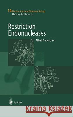
Restriction Endonucleases » książka


Restriction Endonucleases
ISBN-13: 9783540205029 / Angielski / Twarda / 2004 / 443 str.
Restriction Endonucleases
ISBN-13: 9783540205029 / Angielski / Twarda / 2004 / 443 str.
(netto: 764,96 VAT: 5%)
Najniższa cena z 30 dni: 771,08
ok. 22 dni roboczych.
Darmowa dostawa!
Restriction enzymes are highly specific nucleases which occur ubiquitously among prokaryotic organisms, where they serve to protect bacterial cells against foreign DNA. Many different types of restriction enzymes are known, among them multi-subunit enzymes which depend on ATP or GTP hydrolysis for target site location. The best known representatives, the orthodox type II restriction endonucleases, are homodimers which recognize palindromic sequences, 4 to 8 base pairs in length, and cleave the DNA within or immediately adjacent to the recognition site. In addition to their important biological role (up to 10 % of the genomes of prokaryotic organisms code for restriction/modification systems ), they are among the most important enzymes used for the analysis and recombination of DNA. In addition, they are model systems for the study of protein-nucleic acids interactions and, because of their ubiquitous occurence, also for the understanding of the mechanisms of evolution.
Survey and Summary A Nomenclature for Restriction Enzymes, DNA Methyltransferases, Homing Endonucleases and Their Genes.- 1 Introduction.- 2 General Rules.- 3 Details of Types and Subtypes.- 3.1 Types I, II, III and IV.- 3.2 Type I.- 3.3 Type II.- 3.4 Type IIP.- 3.5 Type IIA.- 3.6 Type IIB.- 3.7 Type IIC.- 3.8 Type IIE.- 3.9 Type IIF.- 3.10 Type IIG.- 3.11 Type IIH.- 3.12 Type IIM.- 3.13 Type IIS.- 3.14 Type IIT.- 3.15 Nicking Enzymes.- 3.16 Control Proteins.- 3.17 Type III.- 3.18 Type IV.- 3.19 Hypothetical Enzymes.- 3.20 Homing Endonucleases.- 3.21 Adherence to These Conventions and Updates.- References.- Restriction-Modification Systems as Minimal Forms of Life.- 1 Introduction.- 2 Genomics and Mobility of Restriction-Modification Systems.- 2.1 Genomics.- 2.2 Horizontal Gene Transfer Inferred from Evolutionary Analyses.- 2.3 Presence on Mobile Genetic Elements.- 2.4 Genomic Contexts and Genome Comparison.- 2.4.1 Insertion into an Operon-Like Gene Cluster.- 2.4.2 Insertion with Long Target Duplication.- 2.4.3 Substitution Adjacent to a Large Inversion.- 2.4.4 Apparent Transposition.- 2.4.5 Linkage of a Restriction-Modification Gene Complex with Another Restriction-Modification Gene Complex or a Cell Death-Related Gene.- 2.5 Defective Restriction-Modification Gene Complexes.- 3 Attack on the Host Genome and the Selfish Gene Hypothesis.- 3.1 Post-Segregational Host Cell Killing.- 3.2 Comparison with other Post-Segregational Cell Killing Systems.- 3.3 Selfish Gene Hypothesis.- 3.4 Genomics as Explained by the Selfish Gene Hypothesis.- 4 Gene Regulation in the Life Cycle of Restriction-Modification Systems.- 4.1 Gene Organization.- 4.2 Gene Regulation.- 4.2.1 Restriction Gene Downstream of Modification Gene.- 4.2.2 Restriction Gene Upstream of Modification Gene.- 4.2.3 Modification Enzyme as a Regulator.- 4.2.4 Regulatory Proteins.- 4.2.5 Type I Restriction-Modification Systems.- 4.3 Restriction-Modification Gene Complexes May Be Able to Multiply Themselves.- 5 Intra-Genomic Competition Involving Restriction-Modification Gene Complexes.- 5.1 Two Restriction-Modification Systems with the Same Recognition Sequence Can Block the Post-Segregational Killing Potential of Each Other.- 5.2 Solitary Methyltransferases Can Attenuate the Post-Segregational Killing Activity of Restriction-Modification Systems.- 5.3 Resident Restriction-Modification Systems Can Abort the Establishment of a Similar Incoming Restriction-Modification System.- 5.4 Suicidal Defense Against Restriction-Modification Gene Complexes.- 5.5 Defense Against Invaders by Restriction-Modification Systems.- 6 Genome Dynamics and Genome Co-Evolution with Restriction-Modification Gene Complexes.- 6.1 Some Restriction-Modification Gene Complexes and Restriction Sites Are Eliminated from the Genome.- 6.2 Mutagenesis and Anti-Mutagenesis.- 6.3 End Joining.- 6.4 Homologous Recombination by Bacteriophages.- 6.5 Cellular Homologous Recombination in Conflict and Collaboration with Restriction-Modification Gene Complexes.- 6.6 Selfish Genome Rearrangement Model.- 7 Towards Natural Classification of Restriction Enzymes.- 8 Application of the Behavior of Restriction-Modification Gene Complexes as Selfish Elements.- 9 A Hypothesis on the Attack by Restriction-Modification Gene Complexes on the Chromosomes.- 10 Conclusions.- References.- Molecular Phylogenetics of Restriction Endonucleases.- 1 Discovery and Classification of Restriction Enzymes.- 2 Genomic Context of R-M Systems.- 3 Historical Perspective of Comparative Analyses of Restriction Enzymes: Are They Products of Divergent or Convergent Evolution?.- 4 Crystallography of Type II REases: Exploration of the “Midnight Zone of Homology”.- 5 Homology Between Restriction Endonucleases and Other Enzymes Acting on Nucleic Acids.- 6 Non-Homologous Active Sites in Homologous Structures.- 7 Cladistic Analysis of the PD-(D/E)xK Superfamily.- 8 Identification of PD-(D/E)xK Domains in Other Nucleases and Prediction of Their Position on the Phylogenetic Tree.- 9 In the End, Convergence Wins: Sequence Analyses Reveal Type II Enzymes Unrelated to the PD-(D/E)XK Superfamily.- 10 Evolutionary Trajectories of Restriction Enzymes: Relationships to Other Polyphyletic Groups of Nucleases.- References.- Sliding or Hopping? How Restriction Enzymes Find Their Way on DNA.- 1 Introduction.- 2 Mechanisms of Facilitated Target Site Location by Proteins on DNA.- 2.1 Sliding.- 2.2 Hopping.- 2.3 Intersegment Transfer.- 3 Critical Factors Determining the Efficiency of Target Site Location by Sliding and Hopping Processes.- 4 Sliding or “Hopping” — A Survey of Experimental Data.- 4.1 Structures of Restriction Endonucleases.- 4.2 Accurate Scanning of the DNA for the Presence of Target Sites.- 4.3 Length Dependence of Linear Diffusion.- 4.4 Processivity of DNA Cleavage.- 4.5 DNA Cleavage by a Covalently Closed EcoRV Variant.- 4.6 Cleavage of Topological Connected Plasmid Molecules.- 5 Conclusions.- References.- The Type I and III Restriction Endonucleases: Structural Elements in Molecular Motors that Process DNA.- 1 Energy-Dependent DNA Processing.- 2 Motor Enzyme Architecture of the ATP-Dependent Restriction Endonucleases.- 2.1 Motor Enzyme Motifs in the Type I and III Restriction Endonucleases.- 2.1.1 Gross Organisation of the Type I HsdR Subunits.- 2.1.2 Gross Organisation of the Type III Res Subunits.- 2.1.3 Core Helicase Motifs in ATP Binding and Catalysis.- 2.1.4 The “Q-Tip Helix” — A New Helicase Motif?.- 2.1.5 The DNA Binding Motifs — Family-Specific Deviations.- 2.2 A Motor Enzyme Fold in the Type I and III Restriction Endonucleases.- 2.3 Macromolecular Assembly of the Type I and III Restriction Endonucleases.- 3 Future Directions.- 3.1 Coupling Chemical Energy to Mechanical Motion.- 3.2 Tools for Nanotechnology Rather Than Biotechnology?.- References.- The Integration of Recognition and Cleavage: X-Ray Structures of Pre-Transition State Complex, Post-Reactive Complex and the DNA-Free Endonuclease.- 1 Introduction.- 2 The Pre-Transition State Complex.- 2.1 General Features.- 2.2 DNA Numbering Scheme.- 2.3 Secondary Structure.- 2.4 The EcoRI Kink.- 2.5 Recognition Overview.- 2.6 Sequence-Specific Hydrogen Bonds.- 2.7 Sequence-Specific Interactions Via Bound Water.- 2.8 Sequence-Specific Van der Waals Interactions.- 2.9 Redundancy of Direct Sequence-Specific Interactions.- 2.10 Bound Solvent.- 2.11 “Buried” DNA Phosphate Groups.- 2.12 Solvent-Mediated Contacts to the DNA Bases Flanking the Recognition Site.- 2.13 DNA Minor Groove.- 2.14 Contrasting the 1.85 and 2.7 Å Pre-Transition State Complexes.- 3 The Post-Reactive Complex and the Cleavage Mechanism.- 3.1 EcoRI Endonuclease Crystal Packing.- 3.2 Catalytic Site.- 3.3 The Proposed Cleavage Mechanism.- 3.4 The Integration of EcoRI-Catalyzed DNA Cleavage with Substrate Recognition.- 3.5 Chimeric ß Sheets.- 4 Apo-Eco RI Endonuclease (The Protein in the Absence of DNA).- 4.1 Three Polypeptide Chains in the Asymmetric Unit.- 4.2 Disordered “Arm” Regions.- 4.3 Relation to Crystallographic Packing.- 4.4 Order—Disorder Transition Associated with DNA Binding.- 5 Summary and Conclusions.- References.- Structure and Function of EcoRV Endonuclease.- 1 Introduction.- 2 Structural Characteristics of EcoRV Endonuclease.- 2.1 DNA—Protein Interactions in Specific and Nonspecific Complexes.- 2.1.1 The Specific DNA Binding Mode.- 2.1.2 The Nonspecific DNA Binding Mode.- 2.2 The Structure of the Active Site.- 2.2.1 The Structure in the Absence of Divalent Cations.- 2.2.2 The Location of Bound Divalent Metal Ions.- 2.2.3 The Structure of the Enzyme Product Complex.- 3 Biochemical Characteristics of EcoRV Endonuclease and Structure—Function Relationships.- 3.1 Thermodynamics and Energetics of DNA Binding.- 3.1.1 Specific and Nonspecific Binding to DNA.- 3.1.2 The Energetics of Direct and Indirect Readout Interactions.- 3.2 Chemistry and Kinetics of DNA Cleavage.- 3.2.1 General Aspects of Phosphodiester Hydrolysis.- 3.2.2 Steady State and Rapid Reaction Kinetics.- 3.3 Mechanistic Hypotheses.- 3.3.1 The Role of the Conserved, Catalytic-Motif Residues.- 3.3.2 The Role of Metal Ions and the Need of Conformational Changes in Different Mechanisms.- 3.3.3 Catalytically Active and Inactive Conformational States.- 3.3.4 The Three Metal Ion Mechanism.- 4 Conclusions.- References.- Two of a Kind: BamHI and Bg1II.- 1 Introduction.- 2 BamHI Endonuclease.- 2.1 The Structure of Free BamHI.- 2.2 BamHI Nonspecific Complex.- 2.3 BamHI Specific Complex.- 2.4 The Pre-Reactive Complex of BamHI.- 2.5 The Structure of the BamHI Post-Reactive Complex.- 3 BglII Endonuclease.- 3.1 The Structure of Free BglII.- 3.2 The Structure of BglII Bound to its Cognate DNA Site.- Conclusions.- References.- Structure and Function of the Tetrameric Restriction Enzymes.- 1 Introduction.- 2 Structural Anatomy of the Tetrameric Restriction Enzymes.- 2.1 Monomer Structure.- 2.2 Dimer Arrangement.- 2.3 Tetramer Organization.- 3 Active Sites of the Tetrameric Restriction Enzymes.- 4 DNA Recognition by NgoMIV Restriction Endonuclease.- 5 Possible Model of DNA Recognition by Bse634I/Cfr10I Restriction Endonucleases.- 6 Functional Significance of the Tetrameric Architecture of Restriction Enzymes.- References.- Structure and Function of Type IIE Restriction Endonucleases — or: From a Plasmid that Restricts Phage Replication to a New Molecular DNA Recognition Mechanism.- 1 Introduction.- 2 The General Problem: Type IIE REases Need the Simultaneous Interaction with Two Copies of Their Recognition Sequence for Enzymatic Activity.- 3 Activation of Type IIE REases by Synthetic Oligonucleotide Duplexes.- 4 Activation of Type IIE REases by Cleavage Products and by Non-Cleavable Oligonucleotide Duplexes.- 5 The Enzymes’ Reaction Mechanism — General Aspects and Details.- 5.1 Cooperative Interaction with Two Recognition Sites.- 5.2 Stoichiometry of the Active Enzyme-Substrate Complexes.- 5.3 How Do Type IIE REases Communicate Between Remote DNA Recognition Sites in a DNA Molecule?.- 6 Sequence-Specific DNA Recognition by Type IIE REases.- 7 Domain Organization of Type IIE REases.- 8 Modular Architecture of NaeI and EcoRII and Its Functional Implications.- 9 Reaction Mechanism of Type IIE REases.- 10 Are NaeI and EcoRII Evolutionary Links Between REases, Topoisomerases and DNA Recombinases?.- 11 Does Nature Construct Proteins with New Functions by Shuffling Protein Domains?.- 12 Activation of Type IIE REases for Biotechnological Purposes and for Mapping Epigenetic DNA Modifications.- 13 Final Remark.- References.- Analysis of Type II Restriction Endonucleases That Interact with Two Recognition Sites.- 1 Introduction.- 2 Different Classes of Interactions.- 3 Information from cis Reactions.- 3.1 One-Site/Two-Site Assays.- 3.2 Determining Reaction Mechanism.- 3.3 The Effect of Supercoiling.- 3.4 Catenane Substrates.- 4 DNA Binding and Looping with Sites in cis.- 5 Information from trans Reactions.- 5.1 Kinetic Studies.- 5.2 Binding Studies.- 6 Conclusion.- References.- The Role of Water in the EcoRI—DNA Binding.- 1 Introduction.- 2 Thermodynamics.- 3 Experimental Applications.- 3.1 Equilibrium Competition.- 3.1.1 Osmotic Stress Dependence or Knonsp-sp.- 3.1.2 pH and Salt Dependence of Knonsp-sp.- 3.2 Dissociation Kinetics of EcoRI from Its Specific Site.- 3.2.1 Osmotic Dependence of kd.- 3.2.2 pH and Salt Dependence of kd.- 3.3 Removing Water from an EcoRI—Noncognate DNA Complex with Osmotic Stress.- 3.3.1 Competitive Equilibrium at High Osmotic Stress.- 3.3.2 Dissociation Kinetics of the EcoRI from Noncognate Sites.- 3.4 Other Applications of Osmotic Stress to Restriction Nucleases.- 3.5 Application of Hydrostatic Pressure to Restriction Nucleases.- 4 Summary.- References.- Role of Metal Ions in Promoting DNA Binding and Cleavage by Restriction Endonucleases.- 1 Introduction.- 2 Selection of Metal Cofactors to Promote Endonuclease Activity.- 3 Magnesium Analogs.- 4 Inhibitory Influence of Metal Cofactors.- 5 Illustrative Examples and General Guidelines for Metal-Promoted Endonuclease Activity.- 5.1 EcoRV and EcoRI.- 5.2 PvuII.- 5.3 BamHI.- 6 Other Restriction Endonucleases.- 7 Concluding Remarks.- References.- Restriction Endonudeases: Structure of the Conserved Catalytic Core and the Role of Metal Ions in DNA Cleavage.- 1 Introduction.- 2 Common Structural Attributes of Type II REases.- 3 A Common Catalytic Core with Key Catalytic Sidechains.- 4 Phylogenetic Analysis of REases.- 5 Possible Roles for Divalent Metal Ions.- 6 The One-Two-Three Metal Debate.- 7 Generalizations Regarding REase Catalysis.- 8 One-Metal Catalytic Mechanism: EcoRI, BglII.- 9 Two-Metal Catalytic Mechanisms.- 9.1 EcoRV.- 9.2 BamHI.- 9.3 Tn7 Transposase.- 9.4 T7 Endonuclease I.- 10 Three-Metal Catalytic Mechanism.- 11 “Hypothetical” Active Sites of REase Superfamily Members.- 11.1 MunI.- 11.2 BsoBI.- 11.3 NaeI.- 11.4 MutH.- 11.5 Holiday Junction Resolvases, Hjc.- 12 Prospects for the Future.- References.- Protein Engineering of Restriction Enzymes.- 1 Introduction.- 2 Modifying Contacts of Restriction Enzymes to Their Recognition Sequence.- 2.1 Site-Directed Mutagenesis.- 2.2 In Vivo Selection Systems.- 3 Lengthening of Recognition Sequences.- 4 Changing the Subunit Composition.- 5 Future Directions (In Vitro Evolution).- References.- Engineering and Applications of Chimeric Nucleases.- 1 Introduction.- 2 Engineering of Chimeric Nucleases.- 2.1 Functional Domains in FokI Restriction Endonuclease.- 2.2 Chimeric Nucleases.- 2.3 Zinc Finger Binding and Specificity.- 2.4 Mechanism of Cleavageby Zinc Finger Chimeric Nucleases (ZFN).- 3 Application of Chimeric Nucleases.- 3.1 Stimulation of Homologous Recombination Through Targeted Cleavage in Frog Oocytes Using ZFN: Recombinogenic Repair.- 3.2 Targeted Chromosomal Cleavage and Mutagenesis in Fruit Flies Using ZFN:Mutagenic Repair.- 3.3 Gene Targeting in Human Cells Using ZFN.- 4 Future Experiments.- 4.1 Targeted Chromosomal Cleavage and Mutagenesis of the CCR5 Gene in a Human Cell Line.- 4.2 Targeted Correction of a CFTR Genetic Defect in a Human Cell Line Using ZFN.- 5 Potential Limitations of the Chimeric Nuclease Technology.- 6 Future Outlook.- References.
Restriction enzymes are highly specific nucleases which occur ubiquitously among prokaryotic organisms, where they serve to protect bacterial cells against foreign DNA. Many different types of restriction enzymes are known, among them multi-subunit enzymes which depend on ATP or GTP hydrolysis for target site location. The best known representatives, the orthodox type II restriction endonucleases, are homodimers which recognize palindromic sequences, 4 to 8 base pairs in length, and cleave the DNA within or immediately adjacent to the recognition site. In addition to their important biological role (up to 10 % of the genomes of prokaryotic organisms code for restriction/modification systems!), they are among the most important enzymes used for the analysis and recombination of DNA. In addition, they are model systems for the study of protein-nucleic acids interactions and, because of their ubiquitous occurence, also for the understanding of the mechanisms of evolution.
The present book deals with all aspects of restriction endonucleases including nomenclature, diversity, evolution, genetics, structure and function, mechanism of target site location and DNA recognition, enzymology, protein design, and provides a description of the history of the discovery of and the research on restriction enzymes.
1997-2026 DolnySlask.com Agencja Internetowa
KrainaKsiazek.PL - Księgarnia Internetowa


