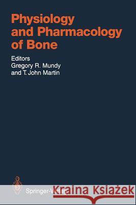
Physiology and Pharmacology of Bone » książka


Physiology and Pharmacology of Bone
ISBN-13: 9783642779930 / Angielski / Miękka / 2011 / 762 str.
Physiology and Pharmacology of Bone
ISBN-13: 9783642779930 / Angielski / Miękka / 2011 / 762 str.
(netto: 383,36 VAT: 5%)
Najniższa cena z 30 dni: 385,52
ok. 22 dni roboczych.
Darmowa dostawa!
Why have another book in the bone-calcium field? When the idea was first raised that this important area of biomedical research should be given more attention in the review literature, we felt that comprehensive texts recently published and in press were directed at more general audiences and did not address in detail some of the important areas of current investigation. As this field has grown and matured, information has accumulated at such a rapid rate that it has become impossible to keep abreast of all areas by reading the original articles in the scientific literature. Accordingly, we believed there was a void which could be filled by a collection of essays on specific topics in which significant progress had recently been made. Thus, this book is meant to be an up-to-date account of specific topics by investi- gators at the forefront of their areas of research. We have tried to review major areas of basic science in the field which have an impact on our understanding of the diseases of bone and mineral. We hope that you will agree that the authors have done an outstanding job, and will find the volume as stimulating to read as we have. San Antonio, TX, USA GREGORY R. MUNDY Melbourne, Australia T. JOHN MARTIN Spring 1993 Contents CHAPTER 1 Calcium Homeostasis A. M. PARFITI. With 12 Figures 1 A. Introduction and Scope. . . . . . . . . . . . . . . . . . . . . . . . . . . . . . . . . . . . . 1 B. Concepts of Homeostasis . . . . . . . . . . . . . . . . . . . . . . . . . . . . . . . . . . . 3 C. Extracellular Fluid Free Calcium: The Controlled Variable. . . . . .
1 Calcium Homeostasis.- A. Introduction and Scope.- B. Concepts of Homeostasis.- C. Extracellular Fluid Free Calcium: The Controlled Variable.- D. Parathyroid Hormone: The Controlling Variable.- I. Afferent Loop: Effects of Calcium on PTH Secretion.- II. Efferent Loop: Effects of PTH on Free Calcium.- III. Short-Term Regulation as a Function of Parathyroid Status.- E. Other Calciotropic Hormones.- I. Calcitriol.- II. Calcitonin.- F. External Balance and Turnover of Calcium.- I. Dietary Intake and Intestinal Absorption.- II. Renal Excretion.- III. Role of Intestine and Kidney in Calcium Homeostasis.- G. Bone and Bone Mineral in Relation to Calcium Homeostasis.- I. Aspects of Bone Remodeling.- II. Role of Bone Remodeling in Calcium Homeostasis.- III. Quiescent Bone Surfaces.- IV. Circulation of Bone, Macro and Micro.- V. Bone Mineral: Composition and Structure.- VI. Movement of Calcium Ions in and out of Bone.- VII. Blood-Bone Equilibrium and Its Homeostatic Function.- VIII. Blood-Bone Equilibrium: Physiochemical and Cellular Mechanism.- H. Integration of Skeletal and Mineral Homeostasis.- References.- 2 Bone Remodeling and Bone Structure.- A. Introduction.- B. Bone Macro- and Microanatomy.- I. Cortical Bone.- II. Cancellous Bone.- C. Bone Remodeling.- I. Quantum Concept of Bone Remodeling.- II. Coupling Phenomenon.- D. Evaluation of Bone Remodeling and Structure.- I. Surface Area Estimates.- E. Evaluation of Bone Resorption.- I. Cortical Bone.- II. Cancellous Bone.- F. Evaluation of Bone Formation.- I. Cortical Bone.- II. Cancellous Bone.- G. Bone Balance.- I. Cortical Bone.- II. Cancellous Bone.- III. Calculation of Activation Frequency.- IV. Calculation of Tissue Level Indices of Turnover.- H. Indices Pertaining to Cancellous Bone Structure.- I. Trabecular Bone Volume.- II. Marrow Space Star Volume.- I. The Bone Resorption Sequence.- I. Cancellous Bone.- II. Cortical Bone.- J. The Bone Formation Sequence.- I. Cancellous Bone.- II. Cortical Bone.- III. The Bone Structural Unit.- K. Bone Remodeling and Bone Loss.- L. Reversible Bone Loss.- I. Cancellous Bone.- II. Cortical Bone.- M. Irreversible Bone Loss.- I. Cortical Bone.- II. Cancellous Bone.- III. Bone Turnover and Bone Loss.- N. Implications for Bone Mass Measurements.- O. Physiological Ageing Processes in Bone: Differences Between Females and Males.- I. Bone Loss in Cortical Bone.- II. Bone Loss in Cancellous Bone.- P. Relationship Between Bone Structure and Bone Strength.- I. Cancellous Bone.- Q. Bone Remodeling in Metabolic Bone Disease.- I. High and Low Turnover Bone Disease.- II. Bone Remodeling in Osteoporosis.- R. Final Remarks and Future Perspectives.- References.- 3 Biology of the Osteoclast.- A. Introduction.- B. Main Morphological Features of the Osteoclast.- C. Structure-Function Relationship.- D. Motility, Attachment, and Establishment of the Bone-Resorbing Compartment.- I. Cytoskeletal Organization.- II. Attachment Apparatus.- 1. Clear Zone.- 2. Podosomes and the Sealing Zone.- 3. Role of Integrins.- 4. Regulation of Bone Resorption and the Attachment Apparatus.- E. Proteins Destined for Export: Biosynthetic and Secretory Functions of the Osteoclast.- I. Lysosomal Enzymes.- II. Nature and Specificity of the Secreted Enzymes.- III. Generation of Oxygen-Derived Free Radicals and Synthesis and Secretion of Other Proteins by the Osteoclast.- F. Cytosolic and Membrane Proteins: Membrane Composition and Ion Transport.- I. Apical Membrane and the Process of Acidification.- II. Role of the Basolateral Membrane and Ion Channels in Acidification, Intracellular pH, and Membrane Potential Regulation.- III. Handling and Regulatory Role of Calcium.- G. Conclusion.- References.- 4 Osteoblasts: Differentiation and Function.- A. Introduction.- B. Mature Members of the Osteoblast Lineage: Definitions.- C. Origin of Osteoblasts.- D. Osteoblast Lineage.- E. Protein Products of Osteoblasts.- F. Factors That Influence Osteoblast Differentiation.- G. Model of Osteoblast Differentiation.- H. Osteoblast Proliferation.- I. Hormone Receptors and Responses of Osteoblasts.- J. Role of Osteoblasts in Intercellular Communication.- I. Osteoclast Activation.- II. Osteoclast Formation.- III. Coupling of Resorption to Formation.- K. Proteinase Production by Osteoblasts.- L. Conclusion.- References.- 5 Cytokines of Bone.- A. Introduction.- B. Nature of the Osteotropic Cytokines.- C. Cell Source of the Osteotropic Cytokines.- D. Interactions Between Systemic Factors and Cytokines.- E. Interactions Between Cytokines.- F. Diseases Associated with Abnormal Cytokine Production.- G. Interleukin-1.- H. Interleukin-1 Receptor Antagonist.- I. Tumor Necrosis Factor and Lymphotoxin.- J. Interleukin-6.- K. Gamma-Interferon.- L. ß2-Microglobulin.- M. Osteoclastpoietic Factor.- N. Colony-Stimulating Factors.- O. Leukemia-Inhibitory Factor (Differentiation-Inducing Factor).- P. Prostaglandins and Other Arachidonic Acid Metabolites.- Q. Transforming Growth Factor ß.- R. Bone Morphogenetic Proteins.- S. Other Bone-Derived Growth Factors.- References.- 6 Hormonal Factors Which Regulate Bone Resorption.- A. Introduction.- B. Parathyroid Hormone.- I. Effects of PTH on Bone Resorption.- II. Effects of PTH on Bone Formation.- III. Signal Transduction Mechanisms for PTH in Bone Cells.- IV. Effects of PTH on Bone Turnover.- V. Effects of PTH on Calcium Homeostasis.- C. 1,25-Dihydroxyvitamin D.- I. Vitamin D Receptor.- II. Effects of 1,25-Dihydroxyvitamin D on Osteoclasts.- III. Effects of Vitamin D Metabolites on Cells of the Osteoblast Lineage.- D. Calcitonin.- E. Amylin.- F. Cortisol.- G. Thyroid Hormones.- H. Estrogens.- References.- 7 Factors That Regulate Bone Formation.- A. Introduction.- B. Platelet-Derived Growth Factor.- C. Heparin-Binding Growth Factors.- D. Insulin-Like Growth Factors and Their Binding Proteins.- E. Transforming Growth Factor Beta.- References.- 8 Mineralization.- A. Introduction.- B. Direct Cellular Control of Mineralization Through Matrix Vesicles.- C. Mechanism of Matrix Vesicle Calcification.- I. Role of Alkaline Phosphatase.- II. Role of Lipids.- III. Constitutive Proteins of Matrix Vesicles.- IV. Biphasic Hypothesis of Mineralization-Crystal Initiation Phase.- V. Biphasic Hypothesis of Mineralization-Crystal Growth Phase.- D. Indirect Cellular Control of Mineralization.- I. Effect of Bone Morphogens on the Calcification Mechanism.- II. Role of Growth Factors in Mineralization.- III. Hormones That Affect Calcification.- IV. Pathological Calcification.- V. Vitamin D.- E. Cellular Regulation of the Composition and Mineralizing Potential of Matrix.- I. Collagen.- II. Proteoglycans and Noncollagenous Proteins of Matrix.- III. Control of Angiogenesis.- IV. Cellular Regulation of the Ionic Milieu at Calcification Sites.- V. Control of pH.- References.- 9 Pathogenesis of Osteoporosis.- A. Introduction.- B. Primary Osteoporosis.- I. Calcium-Regulating Hormones.- 1. Parathyroid Hormone.- 2. 1,25-Dihydroxyvitamin D.- 3. Calcitonin.- II. Systemic Hormones.- 1. Sex Hormones.- 2. Other Systemic Hormones.- III. Local Factors.- 1. Interleukins.- 2. Prostaglandins.- 3. Growth Factors.- IV. Calcium and Other Nutrients.- V. Physical Activity and Life-style.- VI. Alternative Hypotheses.- C. Secondary Osteoporosis.- I. Glucocorticoid-Induced Osteoporosis.- II. Hyperparathyroidism.- III. Hyperthyroidism.- IV. Hypogonadism.- V. Nutritional and Gastrointestinal Disorders.- VI. Renal Disease.- VII. Multiple Myeloma and Other Hematologic Disorders.- VIII. Mast Cells and Heparin.- IX. Osteogenesis Imperfecta.- X. Miscellaneous Therapeutic Agents.- D. Conclusion.- References.- 10 Vitamin D Metabolism.- A. Overview.- B. Dermal Production of Vitamin D3 and Dietary Sources of Vitamin D.- I. Substrate and Chemistry.- II. Ultraviolet Light and Skin Pigmentation.- III. Dietary Sources of Vitamin D.- C. Serum Vitamin D Binding Protein.- I. Ethnic Differences.- II. Binding Affinities and Function.- D. Hepatic 25-Hydroxylation.- I. Microsomal 25-Hydroxylase.- II. Vitamin D Catabolism.- III. 25-Hydroxyvitamin D Bioactivity.- E. Renal 1-Hydroxylation.- I. Proximal Tubule Mitochondrial P450.- II. Regulation of Renal 1-Hydroxylation.- III. Pathophysiology of Renal 1-Hydroxylation.- F. Extrarenal 1-Hydroxylation.- I. Physiological Extrarenal 1-Hydroxylation.- II. Pathological Extrarenal 1-Hydroxylation.- G. Renal and Target Tissue 24-Hydroxylation.- I. Renal 24-Hydroxylation.- II. Target Tissue 24-Hydroxylation.- III. Catabolic Versus Unique Functions of 24,25-Dihydroxyvitamin D.- H. Vitamin D Catabolism.- I. Target Tissues.- I. Bone and Calcium Related.- II. Hematopoietic Cells.- III. Skin and Skin Appendages.- IV. Reproduction and Endocrine Glands.- V. Other Tissues.- J. Vitamin D Receptor.- I. Functional Role.- II. End Organ Resistance.- III. Molecular Biology.- IV. Vitamin D Response Element in Responsive Genes.- K. Genomic and Nongenomic Effects.- L. Summary.- References.- 11 Bisphosphonates: Mechanisms of Action and Clinical Use.- A. Introduction.- B. Chemistry and General Characteristics.- C. Synthesis.- D. Methods of Determination.- E. History of the Development for Use in Bone Disease.- F. Mode of Action.- I. Physicochemical Effects.- II. Effect on Calcification In Vivo.- III. Inhibition of Bone Resorption.- 1. Assessment of Activity.- 2. Activity of Various Bisphosphonates.- 3. Mechanisms of Action of Bone Resorption Inhibition.- 4. Other Effects In Vivo.- G. Pharmacokinetics.- H. Animal Toxicology.- I. Drug Interactions.- J. Clinical Use.- I. Ectopic Calcification and Ossification.- 1. Soft Tissue Calcification.- 2. Urolithiasis.- 3. Dental Calculus.- 4. Fibrodysplasia Ossificans Progressiva.- 5. Other Heterotopic Ossifications.- II. Diseases with Increased Bone Resorption.- 1. Paget’s Disease.- 2. Hypercalcemia of Malignancy and Tumoral Bone Destruction.- 3. Hyperparathyroidism and Other Causes of Hypercalcemia.- 4. Osteoporosis.- III. Other Indications.- K. Adverse Events.- L. Contraindications.- M. Future Prospects.- References.- 12 Paget’s Disease.- A. Introduction.- B. Nature of the Underlying Disease Process.- C. Epidemiology.- D. Direct Studies on the Etiology of Paget’s Disease.- E. Possible Role of Canine Distemper Virus.- F. Properties of the Osteoclast That Might Make It Particularly Susceptible to Persistent Infection with an RNA Virus.- G. Clinical Features.- I. Bones Affected and Extent.- II. Symptoms and Signs.- 1. Features Due to Long-standing Excessive Bony Remodelling.- 2. Features Due to Secondary Arthritis.- 3. Features Due to Pressure on Surrounding Structures.- 4. Neoplasia.- H. Histopathology.- I. Clinical Assessment and Investigation.- J. Treatment of Paget’s Disease.- I. Bisphosphonates (Diphosphonates).- II. Probable Mode of Action of Bisphosphonates in Paget’s Disease.- III. Future Work.- References.- 13 Hyperparathyroid and Hypoparathyroid Bone Disease.- A. Introduction.- B. Parathyroid Hormone Function: Calcium Homeostasis.- C. Classification of Parathyroid Disease.- D. Primary Hyperparathyroidism.- I. Incidence.- II. Diagnosis.- III. Pathogenesis.- IV. Nonskeletal Signs.- V. Skeletal Signs.- 1. Radiology.- 2. Histology.- 3. Bone Mass and Fracture.- VI. Human Parathyroid Hormone.- E. Secondary Hyperparathyroidism.- I. Renal Failure.- 1. Diagnosis.- 2. Pathogenesis.- 3. Skeletal Signs.- II. Vitamin D Deficient Osteomalacia/Rickets.- 1. Pathogenesis and Diagnosis.- 2. Skeletal Signs.- III. Ageing.- 1. Diagnosis and Pathogenesis.- IV. Others.- F. Tertiary Hyperparathyroidism.- G. Hypoparathyroidism.- I. Diagnosis and Pathogenesis.- 1. Nonskeletal Signs.- 2. Skeletal Signs.- References.- 14 Skeletal Responses to Physical Loading.- A. Introduction.- B. Influences on Bone Form.- C. Hierarchy of Functional Control.- D. Nature of the Loading-Related Stimulus.- E. Experimental Studies: In Vivo.- F. Experimental Studies: In Vitro.- G. Loading of Bone Cell Cultures.- H. Implications of Experiments In Vivo.- I. Implications of Experiments In Vitro.- I. Organ Culture.- J. Discussion.- K. Summary.- References.- 15 Parathyroid Hormone: Biosynthesis, Secretion, Chemistry, and Action.- A. Physiologic Actions.- I. Actions in Bone.- 1. Effects upon Osteoclasts.- 2. Effects upon Osteoblasts.- II. Actions in Kidney.- 1. Calcium Reabsorption.- 2. Phosphate Reabsorption.- 3. Other Renal Effects.- B. Biosynthesis and Secretion of PTH.- I. Parathyroid Hormone Biosynthesis.- II. Parathyroid Hormone Gene.- III. Parathyroid Hormone Secretion.- 1. Physiology.- 2. Cellular Mechanisms.- C. Structural Basis of PTH Function.- I. Parathyroid Hormone Binding and Activation Domains.- II. Evolutionary Lessons: Rat and Chicken PTH.- III. Three-Dimensional Structure of PTH.- IV. Lessons from the Structure and Activity of PTHrP.- V. Structure-Activity Relationships for Middle and Carboxyl Regions of PTH.- D. Parathyroid Hormone Receptors.- E. Second Messengers in PTH Action.- I. Cyclic AMP.- II. Other Second Messengers.- III. Physiologic Roles of Different Second Messengers in PTH Action.- IV. Second Messengers in PTH Regulation of Renal Phosphate Transport.- V. Modulation of PTH Signal Transduction.- F. Conclusion.- References.- 16 Calcitonin Gene Products: Molecular Biology, Chemistry, and Actions.- A. Introduction.- B. Calcitonin/CGRP Genes.- I. Structure of DNA.- II. Regulation of Transcription.- III. Tissue-Specific Expression of Messenger RNA.- C. Calcitonin Gene Products.- I. Biosynthesis.- II. Structure and Tissue Distribution.- III. Regulation of Release.- IV. Metabolism.- D. Biological Action.- I. Calcitonin Receptor and Targets.- 1. Receptors.- 2. Biological Targets.- II. Calcitonin Gene-Related Peptide Receptors and Targets.- 1. Receptors.- 2. Biological Targets.- III. Other Gene Products.- E. Clinical Implications.- I. Calcitonin.- II. Calcitonin Gene-Related Peptide.- F. Conclusions.- References.- 17 Parathyroid Hormone-Related Protein: Molecular Biology, Chemistry, and Actions.- A. Introduction.- B. Molecular Biology.- I. Isolation and Cloning.- II. Chromosomal Localization.- III. Genomic Structure.- IV. Regulation of Gene.- V. Recombinant PTHrP.- C. Chemistry.- I. Structure-Activity Relationships.- II. Tertiary Structure.- III. Immunology.- IV. Molecular Processing.- V. Structural Conservation.- D. Actions.- I. Second Messengers and Receptors.- II. Postreceptor Events.- III. Actions on Bone.- IV. Actions on Kidney.- E. Physiological Functions.- I. Parathyroid Hormone Related Protein as an Oncofetal Hormone.- II. Paracrine Agent in Smooth Muscle Relaxation.- III. Parathyroid Hormone Related Protein in Lactating Breast.- IV. Location in Epithelia.- V. Endocrine and Paracrine Roles.- References.- 18 Pathophysiology of Skeletal Complications of Cancer.- A. Introduction.- B. Frequency of the Skeleton as a Site for Malignant Disease.- C. Cancers Which Involve the Skeleton.- D. Favored Skeletal Sites of Malignancy.- E. Complications of the Metastatic Process.- F. Pathophysiology of the Metastatic Process.- I. Properties of Tumor Cells Which Favor Metastasis.- II. Tumor Cell Invasion at the Primary Site.- 1. Adhesion.- 2. Secretion of Enzymes.- 3. Cell Motility.- III. Tumor Cells in the Bloodstream.- IV. Tumor Cell Arrest at the Metastatic Site.- V. Growth Regulatory Factors at Metastatic Sites.- G. Potential Mechanisms for the Metastatic Process in Bone.- I. Chemotactic Factors.- II. Growth Regulatory Factors.- III. Calcium.- IV. Proteolytic Enzymes.- H. Factors Which May Be Involved in Osteolysis.- I. Parathyroid Hormone Related Protein.- II. Transforming Growth Factor a.- III. Transforming Growth Factor ?.- IV. Prostaglandins of the E Series.- V. Parathyroid Hormone.- VI. Vitamin D Sterols.- VII. Platelet-Derived Growth Factor.- VIII. Procathepsin D.- IX. Bone-Resorbing Cytokines.- I. Factors Involved in Osteoblastic Effects.- I. Transforming Growth Factor ?2.- II. Fibroblast Growth Factor.- III. Plasminogen Activator Sequence.- References.- 19 The Proteins of Bone.- A. Introduction.- B. Collagen.- I. Structure and Synthesis of Bone Collagen.- 1. Structure of Collagens.- 2. Type I Collagen Synthesis and Secretion.- II. Disorders of Collagen Synthesis: Osteogenesis Imperfecta.- III. Markers of Collagen Metabolism.- 1. Markers of Type I Collagen Synthesis: Propeptides.- 2. Markers of Collagen Degradation.- C. Gamma-carboxyglutamic Acid Containing Proteins of Bone.- I. Gamma-Carboxy glutamic Acid (GLA).- II. Osteocalcin.- 1. Structure, Biosynthesis, and Tissular Distribution.- 2. Functional Role.- 3. Circulating Osteocalcin.- III. Matrix GLA-Protein.- D. Proteoglycans.- I. Biglycan and Decorin.- 1. Structure.- 2. Distribution.- 3. Properties and Potential Role.- II. CS-PGIII.- III. Other Proteoglycans.- E. SPARC/Osteonectin.- I. Structure.- II. Binding to Hydroxyapatite and to Other Proteins.- III. Tissular Distribution.- IV. Biological Properties and Potential Role.- V. Circulating SPARC/Osteonectin.- F. Bone RGD-Containing Proteins.- I. Bone Sialoproteins.- 1. Osteopontin.- 2. Bone Sialoprotein II.- II. Thrombospondin.- III. Fibronectin.- G. Other Proteins in Bone.- I. Proteases and Protease Inhibitors.- II. Plasma Proteins.- H. Conclusion.- References.- 20 Bone Morphogenetic Proteins.- A. Introduction.- B. Biochemistry and Molecular Biology of the BMPs.- I. In Vivo Assay System.- II. Discovery of Multiple Related Proteins.- 1. Purification from Bone.- 2. Bone Morphogenetic Protein Family.- 3. Recombinant Expression.- 4. Expression of BMPs in Other Tissues.- 5. Chromosomal Localization of the BMP Genes.- C. Activities of Individual BMP Molecules.- I. In Vivo Activities.- II. In Vitro Activities.- III. Bone Morphogenetic Proteins in Embryogenesis.- D. Clinical Utility of BMPs.- I. Clinical Indications.- II. Animal Studies.- 1. Bone-Derived Extracts.- 2. Recombinant Human BMPs.- III. Human Studies with Bone Extracts.- E. Summary.- References.
1997-2026 DolnySlask.com Agencja Internetowa
KrainaKsiazek.PL - Księgarnia Internetowa


