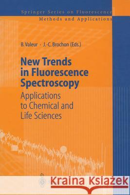
New Trends in Fluorescence Spectroscopy: Applications to Chemical and Life Sciences » książka


New Trends in Fluorescence Spectroscopy: Applications to Chemical and Life Sciences
ISBN-13: 9783642632143 / Angielski / Miękka / 2012 / 490 str.
New Trends in Fluorescence Spectroscopy: Applications to Chemical and Life Sciences
ISBN-13: 9783642632143 / Angielski / Miękka / 2012 / 490 str.
(netto: 192,11 VAT: 5%)
Najniższa cena z 30 dni: 192,74
ok. 16-18 dni roboczych.
Darmowa dostawa!
Fluorescence is more and more widely used as a tool of investigation, analysis, control and diagnosis in many fields relevant to physical, chemical, biological and medical sciences. New technologies con- tinuously emerge thanks to the progress in the design of light sources (e.g. laser diodes), detectors (3D, 4D) and compact ultrafast elec- tronic devices. In particular, much progress has been made in time- resolved fluorescence microscopy (FUM: Fluorescence Lifetime Imaging Microscopy; FCS: Fluorescence Correlation Spectroscopy). Furthermore, the sensitivity now allows one to detect a single mole- cule in the restricted field of a confocal microscope, which actually offers the possibility to study phenomena at a molecular level. The development of new fluorescent probes is still a necessity. In particular, the growing use of lasers implies high resistance to photo- degradation. Fluorescence emission at long wavelengths is also a distinct advantage. Furthermore, in vivo inclusion of new fluorescent aromatic residues in proteins offer new potentialities in biology. of ions and molecules is Fluorescence-based selective detection still the object of special attention. Considerable effort is being made in the design of supramolecular systems in which the recognition event is converted into a fluorescence signal easily detected. New fluorescent sensors for clinical diagnosis and detection of pollutants in atmosphere and water are extensively developed. All these developments justify the regular publication of books giving the state-of-the-art of the methods and applications of fluo- rescence spectroscopy.
1 Historical Aspects of Fluorescence.- 1 Introduction: On the Origin of the Terms Fluorescence, Phosphorescence, and Luminescence.- References.- 2 Pioneering Contributions of Jean and Francis Perrin to Molecular Luminescence.- 2.1 Introduction.- 2.2 Biographical Sketches of Jean Perrin and Francis Perrin.- 2.3 The Perrin-Jablonski Diagram.- 2.3.1 Jablonski Diagram.- 2.3.2 États Métastables — Phosphorescence.- 2.4 Resonance Energy Transfer.- 2.5 Fluorescence Polarization.- 2.6 Concluding Remarks.- 2.7 Bibliographical Notes.- References.- 3 The Seminal Contributions of Gregorio Weber to Modern Fluorescence Spectroscopy.- 3.1 Overview.- 3.2 EarlyYears.- 3.3 Cambridge.- 3.4 Francis Perrin’s Influence.- 3.5 Ph.D. Thesis.- 3.6 Postdoctoral.- 3.7 Sheffield.- 3.8 Intrinsic Protein Fluorescence.- 3.9 Red-Edge Effects.- 3.10 EEM.- 3.11 Brandeis.- 3.12 University of Illinois.- 3.13 Phase Fluorometry.- 3.14 Polarization Revisited.- 3.15 Students, Postdocs and Visitors.- 3.16 Commercialization of Fluorescence.- 3.17 National Laboratories.- 3.18 Honors.- 3.19 Proteins and Pressure.- References.- 2 Fluorescence of Molecular and Supramolecular Systems.- 4 Investigation of Femtosecond Chemical Reactivity by Means of Fluorescence Up-Conversion.- 4.1 Nanosecond and Picosecond Time-Resolved Fluorescence Techniques.- 4.1.1 Phase Modulation Spectroscopy.- 4.1.2 Time Correlated Single Photon Counting.- 4.1.3 Streak Cameras for Time-Domain Measurements.- 4.2 Femtosecond Emission Spectroscopy by Time-Gated Up-Conversion.- 4.2.1 Historical Background of the Time-Gated Up-Conversion Technique.- 4.2.2 Principle of the Time-Gated Up-Conversion Technique.- 4.2.2.1 Phase Matching Conditions.- 4.2.2.2 Quantum Efficiency for Up-Conversion.- 4.2.2.3 Group Velocity Effects.- 4.2.3 Experimental Setup.- 4.3 Time-Resolved Spectroscopy.- 4.3.1 Solvation Processes.- 4.3.1.1 Time-Dependent Fluorescence Stokes Shift (TDFSS) Non-Specific Solvation.- 4.3.1.2 Specific Solvation: Role of the Structure and the Charge of the Probe.- 4.3.1.3 Specific Solvation: Hydrogen Bond Dynamics.- 4.3.1.4 Isotope Effect.- 4.3.1.5 Spectral Narrowing in the 10 ps Time Scale.- 4.3.2 Photoinduced Intramolecular Charge Transfer.- 4.3.3 Intermolecular Electron Transfer.- 4.3.4 Intramolecular Proton Transfer.- 4.3.5 S2?S1 Internal Conversion.- 4.3.6 Biological Systems.- 4.4 Conclusions.- References.- 5 Spectroscopic Investigations of Intermolecular Interactions in Supercritical Fluids.- 5.1 Introduction.- 5.2 Instrumentation.- 5.3 Sample Preparation and Precautions..- 5.4 Selected Applications.- 5.5 Laser Flash Photolysis.- 5.6 Basic Picture Revealed by These Studies.- 5.7 The Future.- References.- 6 Space and Time Resolved Spectroscopy of Two-Dimensional Molecular Assemblies.- 6.1 Introduction.- 6.1.1 Motivation.- 6.1.2 Models.- 6.2 Experimental.- 6.3 Results and Discussion.- 6.3.1 Inhomogeneous Multilayers: RB 18 and ARA.- 6.3.2 Homogeneous Multilayers: SRH+ARA.- 6.3.3 Multilayers of CV18 and ARA or DPPA.- 6.3.3.1 CV 18 in DPPA.- 6.3.3.2 Cd-Arachidate Multilayers.- 6.3.4 Intralayer Quenching of PYR18 by CV18.- 6.4 Conclusions.- References.- 7 From Cyanines to Styryl Bases — Photophysical Properties, Photochemical Mechanisms, and Cation Sensing Abilities of Charged and Neutral Polymethinic Dyes.- 7.1 Introduction.- 7.2 Cyanine Dyes.- 7.2.1 Photophysical Model Mechanisms.- 7.2.2 Complexation Properties.- 7.3 Styryl Dyes.- 7.3.1 Photophysical Model Mechanisms.- 7.3.2 Complexation Properties.- 7.4 Styryl Bases.- 7.4.1 Photophysical Model Mechanisms.- 7.4.2 Complexation Properties.- 7.4.2.1 Donor Acceptor Fluoroionophores.- 7.4.2.2 Donor Acceptor Donor Fluoroionophores.- 7.5 Conclusion.- References.- 8 Phototunable Metal Cation Binding Ability of Some Fluorescent Macrocydic Ditopic Receptors.- 8.1 Introduction.- 8.2 Anthraceno Coronands.- 8.2.1 Free Ligand.- 8.2.2 In the Presence of Metal Cation.- 8.3 Benzeno Coronands.- 8.3.1 BBO5O5.- 8.3.2 0TTO5O5.- 8.3.3 Fluorescence Anisotropy Experiments with BBO5O5.- 8.4 Conclusion.- References.- 3 Fluorescence in Sensing Applications.- 9 The Design of Molecular Artificial Sugar Sensing Systems.- 9.1 Introduction.- 9.2 Fluorescent Monoboronic Acids.- 9.3 Selective Recognition of Saccharides by Diboronic Acids.- 9.4 Introduction of the Concept of PET (Photoinduced Electron Transfer) Sensors.- 9.5 A Glucose Sensor and an Enantioselective Sensor.- 9.6 Conclusion.- References.- 10 PCT (Photoinduced Charge Transfer) Fluorescent Molecular Sensors for Cation Recognition.- 10.1 Introduction.- 10.2 Principles.- 10.3 PCT Sensors Based on the Interaction Between the Bound Cation and an Electron-Donating Group.- 10.3.1 Crown-Containing PCT Sensors.- 10.3.2 Chelating PCT Sensors.- 10.3.3 Cryptand-Based PCT Sensors.- 10.3.4 Calixarene-Based PCT Sensors.- 10.4 PCT Sensors Based on the Interaction Between the Bound Cation and an Electron-Withdrawing Group.- 10.4.1 Crown-Containing PCT Sensors.- 10.4.2 Calixarene-Based PCT Sensors.- 10.5 Conclusion.- References.- 11 Fluorometric Detection of Anion Activity and Temperature Changes.- 11.1 The Two-Component Approach to the Design of a Fluorescent Molecular Sensor.- 11.2 The Use of a [ZnII(tren)]2+ Platform for Anion Recognition and Fluorescent Sensing.- 11.3 Carboxylate Recognition Signalled by Fluorescence Enhancement.- 11.4 The Design of a Molecular Fluorescent Thermometer.- References.- 12 Oxygen Diffusion in Polymer Films for Luminescence Barometry Applications.- 12.1 Introduction.- 12.1.1 Measuring Oxygen Transport.- 12.2 Oxygen Diffusion and Luminescence Quenching.- 12.2.1 Diffusion-Controlled Reactions.- 12.2.2 Quenching and Oxygen Diffusion.- 12.3 Silicone Polymers.- 12.3.1 PDMS.- 12.3.2 Genesee Resins.- 12.4 Poly(aminothionylphosphazenes) (PATP).- 12.5 Modified Poly(aminothionylphosphazenes).- 12.5.1 MSPTP.- 12.5.2 PTHF.- 12.5.3 C4PATP-PTHF Block Copolymers.- 12.5.4 MSPTP-PTHF.- 12.6 Summary.- References.- 13 Dual Lifetime Referencing (DLR) — a New Scheme for Converting Fluorescence Intensity into a Frequency- Domain or Time-Domain Information.- 13.1 Introduction.- 13.2 Theoretical Background.- 13.2.1 Frequency Domain DLR Spectroscopy.- 13.2.2 Time-Domain DLR Spectroscopy.- 13.3 Phosphorescent Standards.- 13.4 Instrumentation.- 13.5 DLR Applications.- 13.5.1 Homogeneous Assays.- 13.5.2 DLR Based Optical Sensors.- 13.5.2.1 Optical Chloride Sensor Based on DLR.- 13.5.2.2 Fiber Optic pCO2 Microsensor Based on DLR.- 13.5.3 DLR Imaging Using Planar Optical pH Sensors.- 13.5.4 Outlook.- References.- 4 New Techniques of Fluorescence Microscopy in Biology.- 14 Two-Photon Fluorescence Fluctuation Spectroscopy.- 14.1 Introduction.- 14.2 Instrumentation.- 14.2.1 Laser.- 14.2.2 Microscope Objectives.- 14.2.3 Microscope, Filters, and Electronics.- 14.3 Autocorrelation.- 14.3.1 Single Species.- 14.3.2 Calibration of the Excitation Volume.- 14.3.3 Comparison of Models.- 14.3.4 Multiple Species.- 14.4 Moment Analysis.- 14.4.1 Comparison Between PCH and Moment Analysis.- 14.5 Conclusions.- References.- 15 Fluorescence Lifetime Imaging Microscopy of Signal Transduction Protein Reactions in Cells.- 15.1 Imaging Protein States by FRET.- 15.2 FRET Imaging by Donor Fluorescence Lifetime.- 15.3 Acceptor Photobleaching in FRET Imaging.- 15.4 Fluorescence Lifetime Imaging Microscopy.- 15.5 Global Analysis and the Population of States.- 15.6 Conclusions.- References.- 16 New Techniques for DNA Sequencing Based on Diode Laser Excitation and Time-Resolved Fluorescence Detection.- 16.1 Introduction.- 16.1.1 The Multiplex Dye Principle and Pattern Recognition.- 16.2 DNA Sequencing in Capillary Gel Electrophoresis by Diode Laser-Based Time-Resolved Fluorescence Detection.- 16.2.1 Semiconductor Lasers as Efficient Excitation Source in the Red Spectral Region.- 16.2.2 Design of Multiplex DNA Sequencing Primers.- 16.2.3 4-Dye-1-Lane Multiplex DNA Sequencing.- 16.3 High-Throughput DNA Analysis.- 16.3.1 Increasing the Speed of Electrophoresis.- 16.3.2 Construction of an Ideal Capillary Array Electrophoresis Instrument (CAE).- 16.3.3 Capillary Array Scanner for Time—Resolved Fluorescence Detection.- 16.3.3.1 Discontinuous Bidirectional Scanning.- 16.3.3.2 Time-Resolved Detection in Parallel Capillaries.- 16.4 Sequencing by Hybridization (SBH).- 16.5 Single Molecule DNA Sequencing in Submicrometer Channels.- References.- 17 The Integration of Single Molecule Detection Technologies into Miniaturized Drug Screening: Current Status and Future Perspectives.- 17.1 Introduction.- 17.2 Theoretical Background of Common Approaches in Single Molecule Analysis (SMA).- 17.2.1 Principles of Fluorescence Correlation Spectroscopy (FCS).- 17.2.2 Autocorrelation Analysis.- 17.2.3 Features and Issues of FCS—Based Screening.- 17.2.4 Photon Counting Statistics: Poisson and Super-Poisson Analysis.- 17.2.5 Photon Counting Histogram (PCH).- 17.2.6 Fluorescence Intensity Distribution Analysis (FIDA).- 17.2.7 Features and Issues of FIDA and PCH.- 17.2.8 Burst Integrated Lifetime (BIFL).- 17.2.9 Features and Issues of BIFL.- 17.3 Conclusion and Outlook.- References.- 18 Picosecond Fluorescence Lifetime Imaging Spectroscopy as a New Tool for 3D Structure Determination of Macromolecules in Living Cells.- 18.1 Time- and Space-Correlated Single Photon Counting (TSCSPC) Spectroscopy and Microscopy.- 18.1.1 DL-System.- 18.1.2 QA-System.- 18.2 EC Biotechnology Demonstration Project: Picosecond Fluorescence Lifetime Imaging as a New Tool for 3 D Structure Determination of Macromolecules in Cells.- 18.2.1 Current State of Knowledge.- 18.2.2 Demonstration Objectives.- 18.2.3 Work Content.- 18.2.4 Role of Partners.- 18.2.4.1 Technology Producers.- 18.2.4.2 Technology Users.- 18.3 Multi-Parameter TSCSPC.- 18.4 Minimal-Invasive Fluorescence Microscopy (MIFM).- 18.5 Living Cells: Fluorescence Dynamics Imaging.- 18.5.1 Fluorescence and Fluorescence Anisotropy Decays of EB-Intercalated DNA in the Cell Nucleus: Collaboration with Maïté Coppey-Moisan (Institut Jacques Monod, Paris).- 18.5.2 GFP-Aggregation, Studied by Fluorescence and Fluorescence Anisotropy Dynamics: Collaboration with Maïté Coppey-Moisan (Institut Jacques Monod, Paris).- 18.5.3 Protein-Protein Interaction: Collaboration with Jürgen Bereiter-Hahn (Goethe University Frankfurt).- 18.5.4 Mitochondria: Fluorescence Dynamics of DASPMI and Rhodamine 700: Collaboration with Jürgen Bereiter-Hahn (Goethe University Frankfurt).- 18.5.5 Chloroplasts: Photosynthesis in Living Plant Cells by Observing Fluorescence Dynamics of the Reaction Centre in Individual Chloroplasts: Collaboration with Hann-Jörg Eckert (TU Berlin).- 18.5.6 The Acquisition of Fluorescence Lifetime Values from Intracellular Sulphonated Aluminium Phthalocyanines Using Confocal Point-Scan and Wide-Field QA Detection [32b] Collaboration with David Phillips (Imperial College, London).- 18.5.6.1 Application of the QA Detector to Obtaining Fluorescence Lifetime Values from Intracellular Sulphonated Aluminium Phthalocyanines.- 18.5.6.2 The Application of Confocal Fluorescence Microscopy in Obtaining Fluorescence Lifetime Values from Intracellular Sulphonated Aluminium Phthalocyanines.- 18.5.6.3 Interpretation of the Results in Terms of Intracellular Phthalocyanine Localisation.- 18.6 Vehicle Micro-Spectroscopy.- References.- 5 Proteins and Their Interactions as Studied by Fluorescence Methods.- 19 About the Prediction of Tryptophan Fluorescence Lifetimes and the Analysis of Fluorescence Changes in Multi-Tryptophan Proteins.- 19.1 Interpreting Fluorescence Changes in Proteins.- 19.2 Determination of the Parameters.- 19.2.1 The Wavelength-Independent Amplitude Fraction ?.- 19.2.2 The Radiative Rate Constant.- 19.3 Analysis of the Meaning of the Different Factors of Q/Q0.- 19.3.1 Heterogeneous Static Quenching or Population Reshuffling (fPR).- 19.3.1.1 Estimation of Microstates of Tryptophan Side Chains.- 19.3.2 The Factor of Pure Dynamic Quenching (fDQ).- 19.4 Examples.- 19.5 Comparison of a System with multiple Fluorophores and Multiple Lifetimes with a System Containing One Fluorophore with Multiple Lifetimes.- 19.5.1 Examples.- 19.6 Conclusion.- References.- 20 Application of Time-Resolved Fluorescence Spectroscopy to Studies of DNA-Protein Interactions and RNA Folding.- 20.1 Introduction.- 20.2 DNA Polymerase Proofreading.- 20.2.1 Detecting the Two DNA Binding Modes of Klenow Fragment.- 20.2.2 Time-Resolved Anisotropy for a Heterogeneous Mixture of Probe Environments.- 20.2.3 Partitioning of Mismatched DNA Substrates Between pol and exo Sites.- 20.2.4 Energetic Contributions of Protein Side Chains to DNA Partitioning.- 20.3 Tertiary Structure Formation in the Hairpin Ribozyme.- 20.3.1 tr-FRET Analysis of the Hairpin Ribozyme.- 20.3.2 Influence of the Interdomain Junction on Ribozyme Folding.- 20.4 Conclusions and Outlook.- References.- 21 Rare Earth Cryptates and TRACE Technology as Tools for Probing Molecular Interactions in Biology.- 21.1 Introduction.- 21.2 Fluorescence and Homogeneous Assays.- 21.2.1 Time Resolved Fluorescence and Rare Earth Complexes.- 21.2.2 Rare Earth Chelates.- 21.3 TRACE Technology.- 21.3.1 Rare Earth Cryptates as a New Type of Fluorescent Label.- 21.3.2 Modulation Processes and Homogeneous Assays.- 21.3.3 Dual Wavelength Detection.- 21.3.4 TRACE Application in Immunoanalysis.- 21.3.5 Kinetic Measurements.- 21.4 TRACE for Probing Molecular Interactions in Life Science.- 21.4.1 Cell Surface Receptor Studies.- 21.4.2 Receptor Tyrosine Kinase Assay.- 21.4.3 Protein-Protein Interactions.- 21.4.4 Protease Assays.- 21.4.5 Applications in Molecular Biochemistry.- 21.4.5.1 Nucleic Acid Hybridization.- 21.4.5.2 Incorporation of TBP Eu3+ Labeled Nucleotides in DNA and RNA.- 21.4.6 “Cassettes” Formats as a Generic Tool.- 21.5 Conclusion.- References.- 22 Tracking Molecular Dynamics of Flavoproteins with Time-Resolved Fluorescence Spectroscopy.- 22.1 Intrinsic Protein Fluorescence.- 22.2 Flavins and Flavoproteins.- 22.3 Flavin as a Fluorescent Probe for Flavoprotein Dynamics.- 22.4 The Intrinsic Flexibility of Proteins.- 22.5 Functionally Important Motions in Flavoenzymes; an Introduction to Glutathione Reductase, Thioredoxin Reductase and p-Hydroxybenzoate Hydroxylase.- 22.6 Current Insights in Flavoprotein Active-Site Dynamics from Fluorescence: the Drive to Higher Time-Resolution, the Revised Interpretation of Heterogeneous Fluorescence Decays, and the Introduction of a New Mechanism for Fluorescence Depolarization.- 22.7 From Ensembles to Single Molecules.- 22.8 Prospects for Studying Conformational Dynamics by Flavin Fluorescence Detection.- References.
1997-2026 DolnySlask.com Agencja Internetowa
KrainaKsiazek.PL - Księgarnia Internetowa


