Joint Imaging Applications in General Neurodegenerative Disease: Parkinson's, Frontotemporal, Vascular Dementia and Autism » książka
Joint Imaging Applications in General Neurodegenerative Disease: Parkinson's, Frontotemporal, Vascular Dementia and Autism
ISBN-13: 9781536194357
Joint Imaging Applications in General Neurodegenerative Disease: Parkinson's, Frontotemporal, Vascular Dementia and Autism
ISBN-13: 9781536194357
(netto: 414,75 VAT: 5%)
Najniższa cena z 30 dni: 421,34
ok. 30 dni roboczych
Bez gwarancji dostawy przed świętami
Darmowa dostawa!
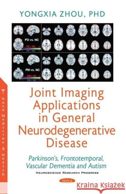
Multiple advanced neuroimaging applications in various neurodegenerative diseases including Parkinson's disease (PD), frontotemporal dementia (FTD), vascular dementia (VaD) and autism spectrum disorder (ASD) are covered in this book. Relatively novel techniques such as integrated PET/MRI and independent component analysis (ICA)-based dual regression (DR) methods were developed to capture multi-level molecular/functional and structural/microstructural as well as high-order inter-network coordination abnormalities. For instance, both PET dopamine transporter and striatal binding ratio reductions in the caudate and putamen were found in PD, consistent with the diffusion tensor imaging (DTI) fractional anisotropy (FA) reduction and fMRI voxel-mirrored homotopic correlation (VMHC) in the substantia nigra (swallow tail sign signature of PD). Furthermore, dopamine storage and pathway labeled with the vesicular monoamine transporter tracer identified decreased densities in the bilateral mesial temporal cortex, caudate, orbitofrontal cortex, left frontal and occipital cortices, consistent with the morphological atrophy, functional connectivity and conductivity deficits in PD. Similarly in FTD patients, the advanced MRI methods such as ICA-DR, VMHC, voxel-based morphometry (VBM) as well as PET tracer for amyloid accumulation and FDG glucose uptake identified typical brain atrophy, structural dis-connectivity, glucose hypometabolism, higher neuropathological burden, lower interhemispheric correlation as well as disrupted intra- and inter-network modulation in the orbitofrontal and anterior temporal cortices together with insular and frontoparietal networks, with the cerebellum and dorsolateral attentional network as typical compensations. Functional and structural abnormalities had further been elucidated in the VaD dependent participants and autistic children. For instance, both lower FA and VMHC, brain atrophy and functional connectivity deficits, demyelination, axonal degeneration and white matter integrity damage in several white matter tracts were present in the dependent compared to independent participants in VaD data cohort. Increased neuronal activity with higher global fractional amplitude of low frequency fluctuation (fALFF) in the conventional and slow-wave sub-band was confirmed with less efficiency of systematic integration in VaD dependent group. Moreover, in ASD compared to controls, regional gray matter volume and cortical thickness in all four brain lobes increased, whereas white matter volume were decreased in addition to the lower temporal, visual and superior frontal but higher inferior and dorsolateral prefrontal cortical functional connectivities exhibited in ASD. The differences in each type of disease could also be revealed with the same imaging method based on either unique region or distinct brain circuit inter-connection, using VMHC, ICA-DR, DTI, VBM, fALFF and graph-theory based small-worldness analysis. In this book, we have developed and generalized conventional and advanced imaging methodologies to several common neurodegenerative diseases. For instance, we have identified the unique imaging signature for each disease type and the underlying neuropathological mechanism connections with conductivity, structural and microstructural connectivity, intra- and inter-network correlation, systematic integration and efficiency analyses. Our objective, comprehensive and confirmative results indicated great potential in utilizing these quantifications for accurate disease classification and staging. With solid imaging evidence, thorough analysis and generalized applications, this book should capture the interests of readers in the broad fields of brain science, disease diagnosis and effective treatment.


