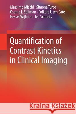
Quantification of Contrast Kinetics in Clinical Imaging » książka


Quantification of Contrast Kinetics in Clinical Imaging
ISBN-13: 9783319646374 / Angielski / Miękka / 2018 / 184 str.
Quantification of Contrast Kinetics in Clinical Imaging
ISBN-13: 9783319646374 / Angielski / Miękka / 2018 / 184 str.
(netto: 191,21 VAT: 5%)
Najniższa cena z 30 dni: 192,74
ok. 22 dni roboczych.
Darmowa dostawa!
This book provides a comprehensive survey of the pharmacokinetic models used for the quantitative interpretation of contrast-enhanced imaging.
Introduction to contrast-enhanced imaging.- Introduction to pharmacokinetic modeling.- Intravascular contrast agents (UCA, blood-pool MRI).- Extravascular contrast agents (MRI, CT, nanodroplets).- Molecular/targeted contrast agents (nuclear imaging and tUCA).
Massimo Mischi (1973) received the M.Sc. degree in Electronic Engineering at La Sapienza University of Rome (Italy) in 1999. In 2004, he received the Ph.D. degree from the Eindhoven University of Technology (TU/e, the Netherlands). In 2007 he became assistant professor and since 2011 he is associate professor at the Electrical Engineering Faculty of the TU/e. Since 2013 he is director of the Biomedical Diagnostics Research Lab of the TU/e (www.bmdresearch.nl), and since 2014 he is director of the Healthcare Research program of the Electrical Engineering Faculty (TU/e). His research focuses on model-based quantitative analysis of biomedical signals and images, spanning from heart and muscle electrophysiology up to ultrasound and magnetic-resonance imaging. He was awarded with the STW VIDI Grant in 2009 and with the ERC Starting Grant in 2011 for his research on contrast-enhanced ultrasound imaging of angiogenesis for cancer diagnostics. Massimo Mischi has (co)authored over 200 publications. He is Senior Member of the IEEE, vice-Chairman of the IEEE EMBS Benelux Chapter, Secretary of the Dutch Society of Medical Ultrasound (EFSUMB Section), and associate board member of the ESUI (Urological Imaging Section of the European Association of Urology). He also serves as associate editor for the IEEE Transactions on Ultrasonics, Ferroelectrics, and Frequency Control and for the Journal BioMedical Engineering and Research (IRBM) published by Elsevier.
Hessel Wijkstra (1955) received the M.Sc. degree in electrical engineering at the Twente University of Technology, Enschede, the Netherlands. He received at the same University the Ph.D. degree with the thesis: ‘The flow pulse response of the ventricular pressure source’. He has been performing bio-medical research in the department of Urology of the Radboud University Hospital, Nijmegen, the Netherlands. Since 2004 he is a faculty member of the department of Urology at the AMC University hospital, Amsterdam, the Netherlands. His main research topic is imaging, in particular contrast enhanced ultrasound, in the diagnosis and treatment of prostate and kidney cancer. Since November 2010 he is part-time professor at the Eindhoven University of Technology focusing on the clinical validation and implementation of contrast enhanced imaging techniques.
Simona Turco received the M.Sc. degree in Biomedical Engineering from University of Pisa (Italy) in 2012, with graduation project focused on laser induced optical breakdown for skin rejuvenation, carried out at the Care & Health laboratories of Philips Research (Eindhoven, the Netherlands). In 2012, she joined the Biomedical Diagnostics (BM/d) Research Lab of the Eindhoven University of Technology (TU/e, Eindhoven) to pursue the Professional Doctor in Engineering (PDEng) diploma in Healthcare Systems Design, working on DCE-MRI dispersion imaging for prostate cancer localization. After obtaining her PDEng diploma in 2014, she became a PhD student at the BM/d research group, where she is currently investigating novel methods for contrast-enhanced and molecular imaging of cancer angiogenesis.
Osama Soliman is qualified from the Medical School of Al-Azhar University, Cairo, Egypt in 1996 Summa Cum Laude. He followed clinical and research training in internal medicine and cardiology at the same institution (1997-2005) and later on (2005-2011) at the Thoraxcenter, Erasmus MC Rotterdam. In March 2011, he received the certificate of completion of training in internal medicine and cardiology. In 2000, he received the M.Sc. (Cum Laude) based on a dissertation entitled “Intravascular ultrasound versus quantitative coronary angiography for the assessment of immediate results of coronary artery stenting”. In 2007, he received the PhD degree from the Erasmus University Rotterdam based on a dissertation entitled “Advanced quantitative echocardiography guiding therapy for heart failure”. He has published more than 120 articles in peer-reviewed journals, practice guidelines, more than 200 abstracts and several book chapters. He is the editor of a book entitled “A practical manual of tricuspid valve diseases”. He is a Fellow of the European Society of Cardiology and the American College of Cardiology. He has been on the Editorial Board of the Journal of the American Society of Echocardiography since 2009 and associate editor of Eurointervention. He is a member of several local and international cardiovascular societies and working groups. He is a well-known expert and director of postgraduate teaching courses in echocardiography and heart failure. His current research focuses on: development of new cardiovascular imaging quantitative methods (imaging); development of advanced novel clinical risk models for end-stage heart failure (heart failure) and clinical trials involving catheter based interventions in structural heart diseases (interventional cardiology).
Ivo Schoots is a staff radiologist at the Department of Radiology & Nuclear Medicine, Erasmus MC, Rotterdam, The Netherlands. He completed his professional radiologic training in 2012 at the Academic Medical Center, Amsterdam after a certified clinical fellowship in abdominal radiology. In 2004 he received his PhD on the ‘crosstalk of coagulation and inflammation in ischemia and reperfusion mechanism’. He received a postdoctoral research fellowship at Harvard Medical School, department of Molecular & Vascular Medicine, BIDMC, Boston (USA). Dr. Schoots is a principle investigator in prostate cancer imaging research. His research is focused on developments in prostate cancer MR imaging and image-guided biopsies, imaging biomarkers, and clinical decision modelling. He is a panel member of the EAU guideline committee on prostate cancer, and participates in international working groups on prostate cancer imaging and image-guided targeted biopsies.
Folkert ten Cate is qualified from the Medical School of Erasmus University, Rotterdam, The Netherlands in 1972. He followed clinical and research training in internal medicine and cardiology at the Thoraxcenter at same institution (1972-1979). In 1979, he received the certificate of completion of training in internal medicine and cardiology. In 1982, he completed a research fellowship on contrast echocardiography at the department of Cardiology, UCLA, Cedars Sinai Medical Center, Los Angeles, USA. In 1978, he received the PhD degree from the Erasmus University Rotterdam based on a dissertation entitled “Asymmetric Septal Hypertrophy: echocardiographic manifestations”. He has published more than 200 articles in peer-reviewed journals, practice guidelines, more than 200 abstracts and several book chapters. He is the editor of several books on contrast echocardiography and valvular heart disease. He is a Fellow of the European Society of Cardiology and the American College of Cardiology. He is a member of several local and international cardiovascular societies and working groups. He is a world renowned expert for his pioneering research in clinical management of hypertrophic cardiomyopathy and development of contrast echocardiography both experimentally and clinically. He is the director of the annual contrast echocardiography congress in Rotterdam since 1996.
This book provides a comprehensive survey of the pharmacokinetic models used for the quantitative interpretation of contrast-enhanced imaging. It discusses all the available imaging technologies and the problems related to the calibration of the imaging system and accuracy of the estimated physiological parameters. Enhancing imaging modalities using contrast agents has opened up new opportunities for going beyond morphological information and enabling minimally invasive assessment of tissue and organ functionality down to the molecular level. In combination with mathematical modeling of the contrast agent kinetics, contrast- enhanced imaging has the potential to provide clinically valuable additional information by estimating quantitative physiological parameters. The book presents the broad spectrum of diagnostic possibilities provided by quantitative contrast-enhanced imaging, with a particular focus on cardiology and oncology, as well as novel developments in the area of quantitative molecular imaging along with their potential clinical applications. Given the variety of available techniques, the choice of the appropriate imaging modality and the most suitable pharmacokinetic model is often challenging. As such, the book provides a valuable technical guide for researchers, clinical scientists, and experts in the field who wish to better understand and properly apply tracer-kinetic modeling for quantitative contrast-enhanced imaging.
1997-2026 DolnySlask.com Agencja Internetowa
KrainaKsiazek.PL - Księgarnia Internetowa


