Physio-Histopathological Changes in the Intestinal Tract of Uromastix » książka
Physio-Histopathological Changes in the Intestinal Tract of Uromastix
ISBN-13: 9783659576669 / Angielski / Miękka / 2014 / 156 str.
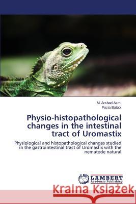
In reptiles Uromastix get preference to study due to have economic importance as its skin used for making fancy articles, its fat used to cure impotence and used as embrocation etc. Among nematode infections Thelandros genus is common in Uromastix. These parasites mainly affect their flesh, skin and fat bodies. Present investigation includes the effect of these parasites on different biochemical parameters of different tissues including glucose, protein, amylase, lipase, alkaline phosphatase and cholinesterase activities and also nucleic acid level of Uromastix for the first time in Pakistan. In this study, its observed that nematodes decreases the DNA rather than other tissues, because nematode parasites present only in intestine of Uromastix but all associated organs of gastrointestinal tract also studied due to their high motility. Histological observations of intestine revealed, hyperplasia, necrosis granulomatous lesion and infiltration of eosinophils, which is represented by photomicrographs. This work is valuable for students of parasitology, teachers and research workers in this field.
In reptiles Uromastix get preference to study due to have economic importance as its skin used for making fancy articles, its fat used to cure impotence and used as embrocation etc. Among nematode infections Thelandros genus is common in Uromastix. These parasites mainly affect their flesh, skin and fat bodies. Present investigation includes the effect of these parasites on different biochemical parameters of different tissues including glucose, protein, amylase, lipase, alkaline phosphatase and cholinesterase activities and also nucleic acid level of Uromastix for the first time in Pakistan. In this study, its observed that nematodes decreases the DNA rather than other tissues, because nematode parasites present only in intestine of Uromastix but all associated organs of gastrointestinal tract also studied due to their high motility. Histological observations of intestine revealed, hyperplasia, necrosis granulomatous lesion and infiltration of eosinophils, which is represented by photomicrographs. This work is valuable for students of parasitology, teachers and research workers in this field.


