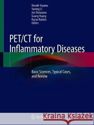
Pet/CT for Inflammatory Diseases: Basic Sciences, Typical Cases, and Review » książka


Pet/CT for Inflammatory Diseases: Basic Sciences, Typical Cases, and Review
ISBN-13: 9789811508097 / Angielski / Twarda / 2020 / 233 str.
Pet/CT for Inflammatory Diseases: Basic Sciences, Typical Cases, and Review
ISBN-13: 9789811508097 / Angielski / Twarda / 2020 / 233 str.
(netto: 651,74 VAT: 5%)
Najniższa cena z 30 dni: 655,41
ok. 16-18 dni roboczych.
Darmowa dostawa!
"A collection of richly illustrated didactic cases, including key images and their interpretation, techniques, and diagnosis ... . Each chapter is complemented by a review of the recent literature. The usefulness of the book may be found mainly in stimulating a greater diffusion of nuclear medical methods ... . in addition to professionals and residents in Diagnostic Imaging, this volume can be of great interest to clinicians who have in their daily experience the diagnosis and treatment of inflammatory diseases." (Pasqualina Sannino and Luigi Mansi, European Journal of Nuclear Medicine and Molecular Imaging, Vol. 48, 2021)
"It is a high-quality textbook written with the cooperation of many doctors and researchers in Japan and China who are active in the field of nuclear medicine. This book will serve as a valuable reference in clinical practice and research for many nuclear medicine physicians and researchers, and I recommend keeping this book on hand as a ready source of information." (Kengo Ito, Annals of Nuclear Medicine, Vol. 34, 2020)
1 Basic science of PET imaging for inflammatory diseases 1.1 Mechanisms of FDG accumulation in inflammation 1.2 FDG PET imaging of inflammation in atherosclerosis 1.3 PET imaging in animal model of neuroinflammation 1 1.4 PET imaging in animal models of neuroinflammation 2 1.5 PET imaging of inflammation in the peripheral system with [18F]FEDAC, a radioligand for translocator protein (18 kDa) 1.6 Role of microglia in neuroinflammation 1.7 Application of preclinical PET imaging for infectious diseases 2 FDG PET/CT in Patients with Inflammation or Fever of Unknown Origin (IUO & FUO) 2.1 Role of FDG PET/CT in the diagnosis of fever or inflammation of unknown origin (FUO/IUO) 2.2 Systematic review and meta-analysis in FDG-PET/CT for inflammatory diseases 2.3 FDG-PET/CT diagnosis of inflammation or fever of unknown origin: a multi-center study in China 2.4 Inflammation of unknown origin 3 FDG-PET/CT for a Variety of Infectious Diseases 3.1 Anti-resorptive agents-related Osteonecrosis of the Jaw (ARONJ) 3.2 Tuberulosis 1 3.3 Tuberculosis 2 3.4 Fungal granuloma 3.5 Infective endocarditis 3.6 Brucella melitensis 3.7 Infective endocarditis 3.8 Splenic ameba 3.9 Liver abscess 3.10 HIV infection 3.11 Traumatic Osteomyelitis 3.12 Hepatic cyst infection in polycystic kidney disease 3.13 Adrenal histoplasmosis 4 Hematological Diseases Mimic Inflammation 4.1 Malignant lymphoma 4.2 Leukemia 1 4.3 Leukemia 2 4.4 Myelodysplastic syndromes 4.5 Hodgkin lymphoma 4.6 Non-Hodgkin lymphoma 4.7 Pulmonary Diffuse Large B-cell Lymphoma Mimicking Lung Abscess Induced by Bronchial Stenosis 5 FDG-PET/CT for Large-Vessel Vasculitis 5.1 Role of FDG PET/CT in diagnosis of inflammation of vessels 5.2 Takayasu arteritis 1 5.3 Takayasu arteritis 2 5.4 Takayasu arteritis 3 5.5 Takayasu arteritis 4 5.6 Takayasu arteritis 5 5.7 Giant cell arteritis 1 5.8 Giant cell arteritis 2 5.9 Giant cell arteritis 3 5.10 Systemic vasculitis 1 5.11 Systemic vasculitis 2 5.12 Aortic graft infection 6 FDG PET/CT for Rheumatic Diseases (Collagen Diseases) 6.1 Role of FDG PET/CT in diagnosis of rheumatic diseases 6.2 Adult-onset Still’s disease 6.3 Polymyositis/Dermatomyositis 6.4 Polymyositis 6.5 Dermatomyositis 1 6.6 Dermatomyositis 2 6.7 Rheumatoid arthritis 1 6.8 Rheumatoid arthritis 2 6.9 Polymyalgia Rheumatica 6.10 Relapsing polychondritis 6.11 Granulomatosis with polyangiitis 1 6.12 Granulomatosis with polyangiitis 2 6.13 Systemic lupus erythematosis 6.14 IgG4 related diseases; Autoimmune pancreatitis and Lymphadenitis 6.15 IgG4RD: Autoimmune Pancreatitis 6.16 IgG4-related cardiovascular disease 7 FDG PET/CT for Sarcoidosis 7.1 Role of FDG PET/CT in diagnosis of cardiac sarcoidosis 7.2 Case 1 7.2 Case 2 7.3 Case 1 7.3 Case 2 7.3 Case 3 7.4 Case 4 7.5 Case 5 8 PET/CT for Neuroinflammation 8.1 Role of TSPO ligands for neuroinflammation 8.2 Alzheimer disease/MCI 8.3 Alzheimer’s disease 8.4 Parkinson’s disease 8.5 Multiple sclerosis 8.6 Neurocysticercosis
Hiroshi Toyama, M.D., Ph.D.,
Department of Radiology,
Fujita Health University
Toyoake, Aichi, Japan
htoyama@fujita-hu.ac.jp
Yaming Li, M.D., Ph.D.,
Department of Nuclear Medicine
The First Hospital of China Medical University
Shenyang, Liaoning, China
ymli2001@163.com
Jun Hatazawa, M.D., Ph.D.,
Specially Appointed Professor,
Joint Research Division for the Quantum Cancer Therapy, Research
Center for Nuclear Physics Osaka University
Professor Emeritus, Osaka University,
Suita, Osaka, Japan
hatazawa@tracer.med.osaka-u.ac.jp
Guang Huang, M.D., Ph.D.,
Shanghai University of Medicine & Health Sciences,Shanghai, China
huang2802@163.com
Kazuo Kubota, M.D., Ph.D.,
Department of Radiology,
Southern TOHOKU General HospitalKoriyama, Fukushima, Japan
kkubota@cpost.plala.or.jp
This comprehensive guide sheds new light on the benefits of FDG PET/CT in diagnosing inflammatory diseases. Although FDG PET/CT offers an invaluable tool for diagnosing inflammatory diseases, the clinical evidence on its application remains limited.
To remedy this gap, each chapter of this book includes detailed descriptions of how FDG PET/CT can be used in connection with a specific inflammatory disease. Further, the authors discusses the precise clinical presentation, including key images andtheir interpretation, techniques and diagnosis. As such, it allows readers to see for themselves how valuable FDG PET/CT is for the diagnosis of cardiac sarcoidosis and aortitis syndrome, as well as rheumatic diseases and neuroimflammation, and the detection of the disease focus of inflammation or fever of unknown origin.
Given its scope, this excellent collection is a valuable resource for radiologists and physicians who are involved in nuclear medicine, as well as cardiologists, cardiovascular surgeons, and rheumatologists.
1997-2026 DolnySlask.com Agencja Internetowa
KrainaKsiazek.PL - Księgarnia Internetowa


