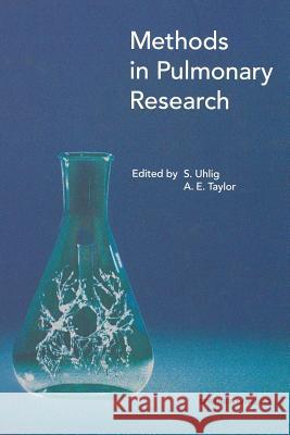
Methods in Pulmonary Research » książka


Methods in Pulmonary Research
ISBN-13: 9783034898034 / Angielski / Miękka / 2012 / 542 str.
Methods in Pulmonary Research
ISBN-13: 9783034898034 / Angielski / Miękka / 2012 / 542 str.
(netto: 191,66 VAT: 5%)
Najniższa cena z 30 dni: 192,74
ok. 16-18 dni roboczych.
Darmowa dostawa!
"Methods in Pulmonary Research" presents a comprehensive review of methods used to study physiology and the cell biology of the lung. The book covers the entire range of techniques from those that require cell cultures to those using in vivo experimental models. Up-to-date techniques such as intravital microscopy are presented. Yet standard methods such as classical short circuit techniques used to study tracheal transport are fully covered. This book will be extremely useful for all who work in pulmonary research, yet need a practical guide to incorporate other established methods into their research programs. Thus the book will prove to be a valuable resource for cell biologists who wish to use organs in their research programs as well biological scientists who are moving their research programs into more cell related phenomena.
Airways.- 1 Measurement of lung function in rodents in vivo.- Spontaneous respiration.- Pulmonary manoeuvres.- Material and equipment.- Lung function laboratory.- Methods.- Preparation and calibration.- Pulmonary function testing.- Examples for applications.- Discussion.- Troubleshooting.- References.- 2 The isolated perfused lung.- Advantages and disadvantages of perfused lungs.- Theoretical background.- Vascular resistance.- Respiratory mechanics.- Material and equipment.- Artificial thorax chamber and ventilation.- Perfusion.- Weight measurement.- Gas exchange.- Methods.- Surgery and setting up the lung.- Criteria for viability.- Cleaning the apparatus.- An example application.- Discussion.- Interpretation of the results.- Constant flow (CFP) versus constant pressure perfusion (CPP).- Negative or positive pressure ventilation.- Choice of perfusate.- Recirculating versus non-recirculating perfusion.- Additional experimental options.- Troubleshooting.- Final comments.- References.- 3 Lung explants.- Material and equipment.- Preparation of culture media.- Preparation of agarose.- Preparation of animals.- Preparation of explants.- Image acquisition.- Variations on this technique.- Applications.- Effects of bronchoconstriction.- Measurements of mucociliary clearance.- Measurements of pulmonary vasculature.- Long term explant culture techniques.- Investigations of protein and gene expression.- Troubleshooting.- Discussion.- Acknowledgements.- References.- 4 Tracheal preparations.- Methods.- Guinea pig tracheal preparations.- Immersion techniques.- Tracheal chain.- Spirally cut trachea.- Zig-zag tracheal strip.- Tracheal tube preparations.- Superfusion techniques.- Electrically stimulated trachea.- Epithelium-denuded trachea.- Conclusion.- References.- Vessels.- 5 Intravital microscopy: Airway circulation.- Materials and equipment.- Microscope.- Video equipment.- Peripheral equipment.- Ventilation.- Solutions.- Methods.- Surgery.- Experimental procedure.- Species differences.- Discussion.- References.- 6 The bronchial circulation.- Importance and role of the bronchial circulation.- Postobstructive pulmonary vasculopathy (POPV) and principles of the techniques.- Material and equipment.- Production of POPV in dogs, rats and guinea pigs: Ligation of the left main pulmonary artery.- In situ perfused LLL preparation.- Morphological assessment of the bronchial and pulmonary vasculature using light microscopy and morphometry.- Methods.- Surgical ligation of the left main pulmonary artery in dogs, rats and guinea pigs.- Canine model.- Rat and guinea pig model.- In situ perfused LLL preparation to measure pulmonary and bronchial vascular flows, pressures and resistances using modified AO and VO and bronchial vascular micropuncture.- Procedure for the in situ perfused LLL preparation.- AO and VO measurements.- Modified in situ perfused LLL preparation for bronchial collateral.- vascular pressure measurements by micropuncture.- Morphological assessment of the bronchial and pulmonary vasculature, using light microscopy and morphometry.- Measurement of pulmonary vascular medial thickness and muscularization in lungs injected with pigmented gelatin-barium mixtures.- Fixation and preparation.- Morphometry.- Assessment of proliferation in the bronchial vasculature.- Bronchial vessel number per airway.- Assessment of bronchial vascular endothelial proliferation using bromodeoxyuridine (BrdU) labeling.- Discussion and troubleshooting.- Production of POPV.- In situ perfused left lower lobar preparation.- Morphological assessment of the bronchial and pulmonary vasculature.- Acknowledgements.- References.- 7 Segmental vascular resistance and compliance from vascular occlusion.- Methods.- The lumped parameter RCR model.- The continuous RC distribution.- More distributed lumped parameter models.- The 3C4R model.- The 3C2R model.- Arterial occlusion in vivo.- Acknowledgements.- References.- Edema.- 8 Experimental and clinical measurement of pulmonary edema.- Definitions.- Lung water filtration and clearance.- Lung water filtration.- Lung water clearance.- Edema formation = filtration ? clearance.- Lung protein filtration and clearance.- Protein filtration.- Protein clearance.- Protein accumulation = filtration ? clearance.- Mechanisms of pulmonary edema.- Material and methods.- Quantifying pulmonary edema formation and clearance in the experimental setting.- Lung microvascular filtration rate.- Isolated lung.- Intact lung.- Lung water clearance.- Lymph flow.- Airway fluid clearance.- Pleural fluid clearance.- Lung water = filtration ?clearance.- Lung weight (isolated lung).- Indicator dilution.- Gravimetry.- Pathology.- Starling equation components.- Microvascular pressure.- Indirect measurements.- Interstitial liquid pressure.- Plasma protein osmotic pressure.- Interstitial colloid osmotic pressure.- Filtration coefficient (Kf,c).- PS.- Sigma (?).- The filtered volume method.- Quantifying pulmonary edema formation and clearance in the clinical setting.- Lung water and edema.- Indicator dilution technique: Extravascular thermal lung volume.- Imaging techniques.- Transthoracic bioimpedance.- Solute filtration: Capillary-alveolar macro-molecule transport.- External radioflux detection.- Positron emission tomography (PET).- Magnetic resonance imaging (MRI).- Edema fluid protein and Bronchoalveolar Lavage fluid.- Starling equation components.- Microvascular pressure.- Small solute PS.- Lung water clearance.- Epithelial permeability: DTPA clearance.- Identifying pulmonary edema and its mechanism in the clinical setting.- Present.- Diagnosis and quantification of pulmonary edema.- Identification of mechanisms.- Identification of an imbalance in Starling forces.- Identification of an altered transvascular permeability.- Future.- Role of edema clearance.- Role of exchange surface area.- Macromolecule transport.- Anatomic distribution.- Conclusion.- References.- 9 Neurogenic inflammation in the airways: Measurement of microvascular leakage.- Material and equipment.- Methods.- Anaesthesia.- Surgery.- Experimental procedure.- Direct electrical stimulation of the vagus nerve.- Chemical stimulants.- Capsaicin.- Bradykinin.- Cigarette smoke.- Sodium metabisulphite.- Other stimulants.- Quantification.- Evans blue dye technique.- Monastral dyes as tracers.- [125?]-albumin.- Application.- Species differences.- Discussion.- Troubleshooting.- Difficulty in cannulating veins.- Difficulty in cannulating arteries.- Poor blood pressure trace.- Acknowledgements.- References.- 10 Intravital microscopy: Surface lung vessels and interstitial pressure.- Material and equipment.- Pipette preparation.- Methods.- Surgery.- Cannulation of left atrium and pulmonary artery.- Preparation of the intact parietal pleural window.- Video imaging analysis.- Experimental procedure.- Application.- Physiological conditions.- Transition to edema.- Lung fluid balance in the newborn.- Mechanical behavior of interstitial matrix.- Video image analysis of the superficial lung structures.- Species differences.- Discussion.- Troubleshooting.- Technical problems.- General drawbacks of the micropuncture technique.- Micropuncture through the intact parietal pleural window.- References.- 11 Lymphatics.- Basic physiology of the lymphatic system.- Methods.- Common technical problems.- Lymph flow rate measurement.- Lymph protein concentration.- Discussion.- Acknowledgements.- References.- Airway liquid.- 12 Evaluation of secretory and transport processes which determine the composition of airway surface liquid.- Methods.- Studies using isolated trachea.- The ferret isolated whole trachea in vitro preparation.- Protocol for stimulating secretions.- Assay for lysozyme.- Albumin transport.- Measurement of potential difference across the tracheal wall.- Ion transport across the airways.- Measurement of the ionic composition of periciliary fluid.- Electrophysiological methods used in the investigation of ion transport in the airways. Measurement of short-circuit current (ISC) and transepithelial resistance (RT).- Acid/base transport.- Acid/base transport across cell membranes of isolated tracheocytes.- Culture of ovine tracheal submucosal gland cells.- Preparation and characterisation.- Methods for studying the effects of secretagogues on lysozyme release from cultured ovine trachea submucosal gland cells.- Electrophysiology.- Troubleshooting.- Conclusions.- Acknowledgements.- References.- 13 Bronchoalveolar lavage.- Methods.- Endoscopic techniques of BAL in man.- Premedication.- Local anaesthesia.- Site of lavage.- Fluid used to perform lavage.- Methods to instil and recover the fluid.- Volumes of fluid to be used.- Recovery.- Should the first aliquot of lavage be processed separately?.- Handling of the harvested lavage material.- Mucus filtration.- Conventional stains.- Membrane filters.- Cytocentrifuge preparations.- Romanovsky stain.- Papanicolaou stain.- Grocott methenamine silver stain-microwave method.- Gram stain.- Iron stain.- In situDNA hybridization.- Immunocytochemical stains.- Procedure (as one example out of a variety of techniques).- Flow cytometry to quantify lymphocyte subsets.- Principle of flow cytometry.- Preparation of samples.- Analysis by immunofluorescence.- Electron microscopy.- Differential cell counts.- Cultures from BAL.- Microbial culture.- Routine culture.- Fungal culture.- Mycobacterial culture.- Viral culture.- Analysis of soluble components of the epithelial lining fluid.- Attempt to quantify lavage material.- Complications of lavage.- Preparation techniques in animals.- References.- 14 Assessment of surfactant function.- In vitro methods for assessment of surfactant function.- The Langmuir-Wilhelmy balance.- Bubbles on a tube: The pulsating bubble surfactometer according to Enhorning.- Captive bubbles.- Microbubble stability.- Adsorption.- The rate of adsorption.- Adsorption characteristics of pulmonary surfactants.- Spreading.- The measurements of adsorption and spreading.- Adsorption.- Spreading.- Measurement of surface tension in situ.- Choosing a test fluid.- Calibration.- Alveolar miropuncture and surface tension in situ.- Influence of surfactant on static lung pressure-volume characteristics.- Animal models for in vivo evaluation of exogenous surfactants.- Preterm newborn animals.- In vivo lung lavage.- Comments.- Acknowledgements.- References.- Cell culture.- 15 Isolation of type II alveolar epithelial cells.- Why do we need to isolate cells for in vitro studies?.- Strategies for isolation of type II cells.- Methods.- Steps in type II cell isolation.- Removal of blood products and alveolar macrophages.- Dissociating lung tissues with digestive enzymes.- Selecting the enzyme combinations.- Recovery of dissociated cells.- Strategies for selective isolation.- Characterization of isolated type II cells.- Morphology.- Surfactant phospholipid profile.- Immunochemical techniques.- Adhesion and culture on different substrata.- Selection of CO2 atmospheric conditions.- Selection of substrata for culture.- Mn++-enhanced technique for pneumocyte isolation.- Reagents.- Type II Cell Isolation.- Discussion.- Limitation of studies of isolated cells.- Alternatives to cell isolation.- Acknowledgments.- References.- 16 Endothelial cells.- Material and methods.- Tissue culture of pulmonary artery endothelial cells.- Equipment/media and chemicals.- Solutions/media preparation.- Macrovascular endothelial cell isolation procedure.- Tissue culture of pulmonary microvascular endothelial cells: Method 1.- Equipment/media and chemicals.- Solutions/media preparation.- Microvascular endothelial cell isolation procedure.- Endothelial cell procurement.- Tissue culture of pulmonary microvascular endothelial cells: Method 2.- Solutions/media preparation.- Microvascular endothelial cell isolation procedure, lung isolation.- Endothelial cell procurement.- Establishment of primary cultures.- Verification of endothelial cells.- Discussion.- Troubleshooting.- References.- Histology.- 17 Studying lung ultrastructure.- Material and equipment.- List of equipment.- Solutions.- Primary fixative for conventional TEM and SEM.- Primary fixative for immunocytochemistry.- Cryoprotectant for infiltration of specimens.- Postosmication for SEM/TEM.- Postfixation for TEM.- Postfixation for SEM.- Methods.- Modes of fixation.- Fixation by airway instillation.- Fixation by vascular perfusion.- Combined chemical/physical fixation for immunocytochemistry.- Fixation by immersion into a chemical fixative.- Sampling of tissue blocks.- Processing and embedding of tissue blocks.- For conventional LM.- For conventional TEM.- For TEM based immunocytochemistry.- For SEM modified according to an OTOTO method.- Sampling of micrographs.- Discussion.- Selecting the mode of fixation.- Selecting the mode of application.- Selecting the fixing agent.- Selecting the mode of tissue processing.- Stereological analysis.- Interpretation.- Acknowledgements.- References.- 18 Autoradiography in the lung.- Materials and equipment.- Methods.- Slide preparation.- Slide cleaning and subbing.- Preparation of chrome alum/gelatin solution.- Application.- Preparation of emulsion-coated coverslips.- Safelight illumination.- Emulsion coating of coverslips.- Tissue preparation.- Tracheal and bronchial tissue.- Peripheral lung tissue.- Cutting frozen sections.- Radioligand binding procedures.- Attachment of emulsion-coated coverslips.- Emulsion-dipped autoradiograms.- Film autoradiograms.- Exposure of autoradiograms.- Development and staining of autoradiograms.- Preparation for development and tissue staining.- Processing solutions.- Photographic development.- Tissue staining and mounting.- Materials.- Procedure.- Chemography controls.- Discussion.- Controls.- Choosing the appropriate autoradiographic method.- Emulsion-dipped preparations.- Emulsion-coated coverslip preparations.- Film imaging.- Microscopy.- Photography.- Image analysis.- References.- Further methods.- 19 Application of aerosols.- Methods.- Physical characterization of aerosols.- Methods of aerosol generation.- Methods of aerosol measurement.- Exposure methodology.- References.- 20 Cryopreservation of human pulmonary tissues.- Mechanisms of freezing injury.- Cryoprotective agents and cryomedia.- Freezing procedure and storage temperature.- Thawing procedure.- Methods, material and equipment.- Cryopreservation.- Freezing procedure.- Temperature and sample handling during storage.- Thawing procedure.- Inventory control.- Post-thaw functional recovery.- Conclusion.- Safety recommendations.- References.- Appendix I. Physiological data of various mammalian species.- References.- Appendix II. List of suppliers.
Stefan Uhlig, Dipl.-Volkswirt, ist Unternehmensberater mit dem Schwerpunkt Sanierung und Krisenmanagement mittelständischer Betriebe. Zudem ist er Sachverständigengutachter im Auftrag deutscher Gerichte zu betriebswirtschaftlichen Problemen und leitet Seminare der IHK zu seinen Schwerpunkten.
1997-2026 DolnySlask.com Agencja Internetowa
KrainaKsiazek.PL - Księgarnia Internetowa


