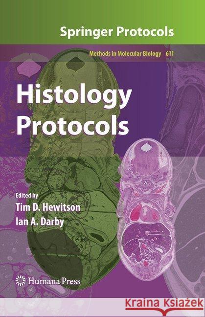
Histology Protocols » książka


Histology Protocols
ISBN-13: 9781493956944 / Angielski / Miękka / 2016 / 229 str.
Histology Protocols
ISBN-13: 9781493956944 / Angielski / Miękka / 2016 / 229 str.
(netto: 460,04 VAT: 5%)
Najniższa cena z 30 dni: 462,63
ok. 16-18 dni roboczych.
Darmowa dostawa!
This book provides molecular biologists with the basic histochemical techniques and histologists with the molecular techniques necessary to realize the potential of their resource. Authoritative and cutting-edge, the book covers a wide range of techniques.
From the reviews:
"This slim volume aims to provide molecular biologists with the basic histochemical techniques and histologists with the molecular techniques to realise their potential of their resource. ... I enjoyed reading this book ... . this book is not particularly expensive and is so comprehensive and well written that I would certainly recommend it to anyone thinking of setting up a research project but uncertain what sort of method to use. It would also be a valuable asset to any departmental library ... ." (Anne Patricia Campbell, Bulletin of the Royal Society of Pathologists, Issue 154, April, 2011)
"Reports the basic techniques of immunohistochemistry and molecular biology used to optimize resources and obtain accurate results. ... This book is recommended to students and researchers using the laboratory techniques on histology described. Updated protocols are described by renowned scientists in the area using understandable language while details of possible interference are given for each technique. Tim D. Hewiston and Ian A. Darby have undertaken several studies on histology, many of which were conducted in collaboration with the authors contributing to this book." (Rui Curi, Brazilian Journal of Pharmaceutical Sciences, Spring, 2011)
"This volume in the Methods in Molecular Biology series provides a compendium of proven protocols for modern histology. ... Written for basic science and clinical researchers interested in mapping the location and distribution of biomarkers, the book also will be appreciated by lecturers and students specializing in biomedical technology, histology, and clinical laboratory medicine. ... The book is well written and carefully edited, and will be a welcome resource for all basic science and clinical researchers studying modern cell biology." (Bruce A. Fenderson, Doody's Review Service, June, 2010)
Part I: Tissue Processing 1. Tissue Preparation for Histochemistry: Fixation, Embedding, and Antigen Retrieval for Light Microscopy Tim D. Hewitson, Belinda Wigg, and Gavin J. Becker 2. An Optimized RNA Extraction Method from Archival Formalin Fixed-Paraffin Embedded Tissue Joon-Yong Chung and Stephen M. Hewitt 3. Laser Capture Microdissection and Pressure-Catapulting for the Analysis of Gene Expression in the Renal Glomerulus Amanda J. Edgley, Renae M. Gow, and Darren J. Kelly Part II: Staining Techniques 4. Immunofluorescence Detection of the Cytoskeleton and Extracellular Matrix in Tissue and Cultured Cells Josiane Smith-Clerc and Boris Hinz 5. Double Immunohistochemistry with Horseradish Peroxidase and Alkaline Phosphatase Detection Systems Vincent Sarrazy and Alexis Desmoulière 6. Retrogradely-Transported Neuronal Tracers Combined with Immunohistochemistry Using Free Floating Brain Sections Emilio Badoer 7. High-Pressure Freezing, Chemical Fixation, and Freeze-Substitution for Immuno-Electron Microscopy Christian Mühlfeld 8. Lectin Histochemistry for Light and Electron Microscopy Su Ee Wong, Catherine Winbanks, Chrishan S. Samuel, and Tim D. Hewitson 9. Duplex In Situ Hybridization in the Study of Gene Co-Regulation in the Vertebrate Brain Raphael Pinaud and Jin K. Jeong 10. Special Stains for Extracellular Matrix Andréa Monte-Alto-Costa and Luís Cristóvão de Moraes Sobrino Porto 11. Active Staining of Mouse Embryos for Magnetic Resonance Microscopy Alexandra Petiet and G. Allan Johnson 12. Immunohistochemical Detection of Tumor Hypoxia Richard J. Young and Andreas Möller 13. In Situ Localization of Apoptosis Using TUNEL Tim D. Hewitson and Ian A.Darby Part III: Imaging Techniques 14. Use of Confocal Microscopy for 3-Dimensional Imaging of Neurons in the Spinal Cord Martin Stebbing, Simon Potocnik, Pinglu Ye, and Emilio Badoer 15. High Resolution Confocal Imaging in Tissue Verena C. Wimmer and Andreas Möller 16. Software-Based Stacking Techniques to Enhance Depth of Field and Dynamic Range in Digital Photomicrography Jörg Piper 17. Image Analysis and Quantitative Morphology Carlos Alberto Mandarim-de-Lacerda, Caroline Fernandes-Santos, and Marcia Barbosa Aguila
With much of what we know about the pathogenesis of disease coming from the systematic and careful study of histological material, histology has proven to be an incredibly valuable area of study. In Histology Protocols, expert researchers in the field contribute detailed methods involving the preparation of tissue, providing an overview of fixation, embedding, and processing, along with the required techniques for the retrieval of RNA from histological sections, followed by sections on routine and specialist histological staining techniques, including immuno-, lectin, and hybridization histochemistry, histological staining, as well as specific methods for the in situ identification of hypoxia and apoptosis, and details on the advances in imaging and image analysis. Written in the highly successful Methods in Molecular Biology™ series format, chapters include introductions to their respective topics, lists of the necessary materials and reagents, step-by-step, readily reproducible laboratory protocols, and notes on troubleshooting and avoiding known pitfalls.
Authoritative and cutting-edge, Histology Protocols will provide molecular biologists with the basic histochemical techniques and histologists with the molecular techniques necessary to realize the potential of their resource.
1997-2026 DolnySlask.com Agencja Internetowa
KrainaKsiazek.PL - Księgarnia Internetowa


