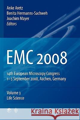
EMC 2008: Vol 3: Life Science » książka


EMC 2008: Vol 3: Life Science
ISBN-13: 9783642098970 / Angielski / Miękka / 2010 / 407 str.
EMC 2008: Vol 3: Life Science
ISBN-13: 9783642098970 / Angielski / Miękka / 2010 / 407 str.
(netto: 766,76 VAT: 5%)
Najniższa cena z 30 dni: 771,08
ok. 16-18 dni roboczych.
Darmowa dostawa!
Proceedings of the14th European Microscopy Congress, held in Aachen, Germany, 1-5 September 2008. Jointly organised by the European Microscopy Society (EMS), the German Society for Electron Microscopy (DGE) and the local microscopists from RWTH Aachen University and the Research Centre Julich, the congress brings together scientists from Europe and from all over the world. The scientific programme covers all recent developments in the three major areas of instrumentation and methods, materials science and life science."
Life Science.- Cryoelectron microscopy: from molecules to systems.- Progress in High-resolution Scanning Probe Microscopy.- Analysis of macromolecules and their supramolecular assemblies.- Modular organization of RNase P.- Molecular anatomy of the human pathogen Leptospira interrogans.- Unveiling conformational changes of biological molecules using multiscale modeling and multiresolution experiments.- Nicolas Boisset: In memoriam.- Cryo TEM-based 3D reconstruction of the recombinant expressed human zinc peptidase Meprin ?.- Origin melting by SV40 Large T antigen-new insights from 3D-EM-MLF3D classification method.- Sequence analysis and modelling of the two large subunits of Phosphorylase Kinase.- Cellular Uptake of Polymer Nanoparticles Imaged by Electron Microscopy.- 9 Å cryo-EM structure and molecular model of a gastropod hemocyanin didecamer (KLH1) reveals the architecture of the asymmetric collar.- Microstructure of model systems for sauces based in polysacharides observed by Cryo-SEM.- Three-dimensional architecture of outer- and inner-dynein arms in flagella revealed by cryo-electron tomography and single particle analysis.- Structural and functional studies of rabbit skeletal muscle Phosphorylase Kinase.- Single-particle analysis of the Cdc6/Orc1, archaeal DNA replication initiator protein.- A high-throughput acquisition toolbox in Matlab for cryo-electron microscopy studies.- Architecture of bacterial glutamate synthase complexe using hybrid approaches.- 3D Structure of DNA Repair Macromolecular Complexes Participating in Non-Homologous End-Joining (NHEJ).- Quaternary structure of recombinant human meprin ?, a zinc peptidase of the astacin family.- Allosterism of Nautilus pompilius hemocyanin as deduced from 8 Å cryo-EM structures obtained under oxy and deoxy conditions.- High resolution structure of a 6 MDa protease by xray-crystallography and cryo-EM.- Cryo-TEM of liquid crystalline particles: Application of materials science techniques to study liquid crystalline particle structures.- Structural basis for the concerted integration of HIV-1 DNA in the human genome, role of the human cofactor LEDGF/p75.- Titan Krios: Automated 3D imaging at ambient and cryogenic conditions.- Self assembly and phase behaviour of new sugar based gemini amphiphiles.- Peptide-nanotube formation by lysine based lipoamino acids.- Advances in biological microanalysis using event streamed spectrum imaging and programmed beam acquisition.- 3D-correlation-averaging for membrane-protein-crystals.- 8 Å cryo-EM structure of the giant hemoglobin from the planorbid snail Biomphalaria glabrata.- Structure of Pex5p and Pex5-20 complexes in the yeast Hansenula polymorpha. Pex20p causes a conformational change upon binding to Pex5p tetramers involved in peroxisomal protein transport.- Structure of OprM-MexA interacting complex revealed by cryo electron tomography.- Cell Structure and Dynamics.- Pleiomorphic viruses revealed by cryo tomography: the structure of coronaviruses.- Combined structural and chemical analysis of unique anammox bacteria that contain a prokaryotic organelle.- Structural and functional considerations on the 3-D organization of the fenestrated cytoplasm of hepatic endothelial cells.- Molecular architecture of the presynaptic compartment studied by cryo-electron tomography.- Synapses in high pressure frozen Caenorhabditis elegans.- High resolution EM-tomography of a whole vitrified cell.- Evaluation of beam damage in catalase crystals observed in vitrified sections.- Urothelial fusiform vesicles are formed in post-Golgi compartment.- Localisation of GFP and GFP tagged PKD2 in cultivated pancreatic cancer cells using high-pressure freezing.- Cytomegalovirus membrane envelopment revisited — a STEM-tomography study based on high-pressure freezing and freeze substitution.- Electron tomography & template matching of biological membranes.- Correlated Microscopy: From Dynamics to Structure.- 3DEM-analyses of Golgi apparatus subunits during physiologic and pathologic reorganizations.- Visualization of the 80s ribosome in situ using cryo electron tomography of vitreous sections.- Three-dimensional analysis of the intermediate filament network using SEM-tomography.- The apical vesicles of the tuft cells in the main excretory duct of the rat submandibular gland by EFTEM-TEM tomography.- (S)TEM Dual Axis Tomography.- STEM tomography of high-pressure frozen cell monolayers.- Multimodality of pericellular proteolysis in cancer cell invasion.- Photonic crystal type nanoarchitectures in butterfly wing scales.- Iridovirus-like viruses of Lacerta monticola from Serra da Estrela, Portugal.- Structural and ultrastructural investigations on the in vitro effects of plant extracts in chicken and rabbit whole blood cultures.- Fine structural characterization of cloned human endogenous retrovirus HERV-K113.- The procaine hydrochlorate effect onto the corpuscular anthocyans from the vacuolar sap of different plant cells.- Comparative anatomical and ultrastructural investigations on normal and vitroplantlets leaves from Pistia stratiotes L..- Autophagy of mitochondria in brown adipocytes of chemically thyroidectomised rats.- Ultrastructural studies of immune organs in antigen primed chickens treated with vegetal extractions.- Structural features of HIV-1 immunologic activation.- Functions of actin-binding proteins in the cell nucleus.- Ultrastructural mechanisms of entamoebae movement system.- White adipocytes transdifferentiation into brown adipocytes induced by triiodothyronine.- Ultrastructural studies of the tegument of cestodes (Platyhelminthes): phylogenetic implications.- Crystallization stages of the CaCO3 deposits in the earthworm’s calciferous gland.- Scanning electron microscopic analysis of heavy metal resistant microorganisms.- Characterization of mouse embryoid bodies by Scanning Electron Microscopy (SEM) and Confocal Laser Scanning Microscopy (CLSM).- Stage-dependent localization of mitochondrial DNA during the cell cycle of Euglena gracilis Z by immunogold electron microscopy.- The virus-inducted structures in RNA virus infected macrophages.- Surface layers of ore-leaching Bacteria and Archaea.- Structure of Bacillus anthracis spores analyzed by CEMOVIS.- Insights into the role of pUL71 during HCMV morphogenesis.- Fine structure of the genital chamber of female Chrysomya megacephala (F.) (Diptera: Calliphoridae).- Ultrastructure of the testes and spermatozoa of the blow fly Chrysomya megacephala (F.) (Diptera:Calliphoridae).- Biosynthesis of cellulose in red algae.- Light and electron microscopy aspects of the glandular sessile hairs from the vitroplantlet leave of Drosera rotundifolia.- Ultrastructure of selected Euglena species in relation to their taxonomy.- Confocal Microscopy Reveals Molecular Interactions Between DNA-binding Drugs and Chromatin in Live Cells.- Fluorescence microscopy methods for measuring the mobility and stability of molecules in 3-D samples.- Phasor-FLIM analysis of FRET for homotypic and heterotypic non-covalent interactions: stimulated membrane receptors in live cells.- In vitro immunofluorescence and ultrastructural analysis of the expression of noncollagenous matrix proteins in human pulpal cells induced by mineralizing factors.- Differentiation-dependent Golgi fragmentation in the bladder urothelial cells in vitro.- Monitoring Mitochordria Dynamics in Living Cell System by Confocal Microscopy.- The freeze-fracture replica immunolabeling technique for the study of lipid droplet biogenesis and function.- Dynamics of epigenome duplication, accessibility and translation.- Controlled light exposure microscopy reveals telomeric microterritories throughout the cell cycle.- Calibration in fluorescence correlation spectroscopy for measurements of stem cell differentiation kinetic.- Spectral methods for cells and particles identifications.- 3D realistic visualization of supramolecular assemblies.- Heterosis in Arabidopsis thaliana: Structural aspects in mature and germinating seeds from hybrid and parental lines.- Colour visualization of red blood cells in native smears by the new method reflected light microscopy.- In vitro culture of Trigonella foenum-graecum plantules and their anatomic characterization.- Live cell imaging by SEM-hosted X-ray microscope in “water window” energy range.- Microscopic anatomy and mineral composition of cuticle in amphibious isopods Ligia italica and Titanethes albus (Crustacea:Isopoda).- In vivo imaging and quantification of the continuous keratin filament network turnover.- Microscopy advances in the life sciences.- Necrotic cell death, a controlled way of cellular explosion.- Dissecting mitochondrial protein distributions using sub-diffraction resolution fluorescence microscopy.- High Thoughput, High Content Tissue Cyotometry.- Focus on the vascular wall: Imaging of large arteries using two-photon microscopy.- High resolution confocal Ca2+ imaging of the pulmonary neuroepithelial body microenvironment in lung slices.- Two photon excitation microscopy and SHG imaging as a tool for visualisation of type I and type II collagen, and their use in tissue engineering.- Testing calibration standards for confocal and two-photon microscopy.- Distribution of the voltage-gated delayed-rectifier K+ subunits, Kv1.1 and Kv1.2, in the adult murine enteric nervous system.- Observation of Ventricular Myocyte Morphology in Long Term Culture using High Resolution Confocal Imaging.- A magnetic field enhances the inmunofluorescence signal in a lymphocytic cell.- Novel 515 nm DPSS-laser brings excitement in life cell imaging.- Confocal laser scanning microscopy study on human fissure caries stained with alizarin red.- Non-invasive skin tissue characterization using non-linear spectral imaging microscopy.- Force Spectroscopy reveals mechanical structure of capsids.- Manipulating and imaging molecular motors with optical traps, single-molecule fluorescence and atomic force microscopy.- Mechanical stability of ethanol-induced A-DNA.- Manipulation of the mechanical properties of a virus by protein engineering.- A single-molecule mechanical assay to study DNA replication coupled to DNA unwinding.- Generation of multiple DNA lesions at subnuclear resolution by multi-photon irradiation.- Light and electron microscopy of phagosomes.- The Use of Electron Microscopy in the Diagnosis of Human Microsporidial Infection — the Manchester, UK, Experience.- Overview of the impact of biological microanalysis in health and disease.- Immunolocalization of FOXP3 in HCV-infected liver biopsies. Preliminary observations..- Ultrastructural and Intracellular Elemental Composition Analysis of Human Hematopoietic Cells During Cold Storage in Preservation Solutions.- Reconstitution and morphological characterisation of an original Human Endogenous Retrovirus.- Cell death in human articular chondrocyte: an ultrastructural study in micromass model..- Pathomorphological diagnostic of paraffin embedded versus epon embedded cardiac tissues with transmission electron microscope analysis.- Interaction of carbon nanotubes with macrophages: STEM and EELS approach.- The ultastructural effect of nitrogen mustard gas on rat brain and therapeutic value of proanthocyanidin.- Assembly and maturation of pestiviruses.- Analysis of radiation-induced bystander effects using high content screening.- Electron microscopic investigations on normal and dexamethasone applied rat placentas.- Changes in dental enamel birefringence after CO2 laser irradiation through fluoride gel -a pilot study.- SEM and light microscopic examination and morphometric analysis of temporal changes in intimal and medial connective tissue component of human carotid arteries.- Ultrastructure of human amniotic membrane covering villous chorion and smooth chorion in preeclampsia.- Changes in Intracellular Sodium, Chlorine, and Potassium Content in Hematopoietic Cells after Hypotermic Storage.- Requirements for a bright future of electron microscopy in the rapid laboratory diagnosis of infectious diseases.- Changed thickness of zona pellucida as a result of stimulated ovulation.- Mozaic microscopy of pancreatic islets.- Microscope history Database.- Interaction of H-Ras Transformed Cell Line with Folic Acid Modified Magnetic Nanoparticles and Detection by Transmission Electron Microscope.- Sedimentation of suspensions for diagnostic thin section electron microscopy.- Evaluation of rofecoxib administration influence on ultrastructural image of the kidney.- Microstructural changes produced by Pulsed Electric Fields in liquid whole egg studied by Transmission Electron Microscopy (TEM).- Microstructural changes in dough treated by Glucose oxidase (GOX) and Transglutaminase (TG), studied by Scanning Electron Microscopy (SEM).- Cytoplasmic K/Na balance in the cardiomyocyte of young and old rats.- Hyperspectral imaging — a novel concept for marker free chromosome characterization.- Poxviruses: Morphogenesis of an Orthopoxvirus (CPXV) and an Avipoxvirus (FPXV) in host tissue.- Ultrastructural alterations of methotrexate in mouse kidney.- Ultrastructural gadolinium evidence in skin of a patient with Nephrogenic Fibrosing Dermopathy.- Nephrotoxicity induced by inorganic Hg(II) and Pb(II): a microscopic and biochemical in vitro study.- Tissue distribution of peroxisomes in zebrafish.- An analysis of the ultrastructure of matured HIV.- Trichinella spiralis and Trichinella britovi cuticle and hypodermal glands Ultrastructure.- Ultrastructural analysis of lysosomal storage diseases: effects of therapy.- Sample preparation and identification of molecular targets.- Immunolabeling for Electronmicroscopy.- Molecular Organisation of Cadherins in Intercellular Adhesion Junctions by Cryo-Electron Tomography of Vitreous Sections.- Ultrastructural observations of indium in the lactating mammary gland cells.- An active mechanism flanks and modulates the export of the small ribosomal subunits.- New methods for micro-domain detection in bacterial sacculi.- Subcellular localization of Myogenic Regulatory Factors along skeletal muscle development.- Immunocytochemical Strategies: LR Resins or Lowicryl, Gold or Peroxidase — Which is Better?.- Elemental Analysis in Electron Microscopy for Medical Diagnostics.- Synaptic localization of KCa1.1 potassium channels in central neurons revealed by postembedding immunogold and SDS-digested freeze-fracture replica labelling.- Ultrastructural studies of rod photoreceptor nuclei from SCA7 mouse.- Application of electron energy loss spectroscopy for heavy metal localization in unicellular algae.- Localization of “flagella” proteins in the Archaeon Ignicoccus hospitalis.- Detection of gold nanoparticles by Autrata YAG detector in FE SEM working in cryo mode.- Correlative cryo-fluorescence and electron microscopy.- The effect of hypometabolizing molecules on transcription as shown by a new two-step pulse-chase method.- Cryofixation and Freeze-Substitution Combined with Tokuyasu Cryo-section Labelling.- The Drosophila active zone architecture: combining confocal, STED and transmission electron microscopy.- Distribution and function of amorphous CaCO3 and calcite within the tergite cuticle of terrestrial isopods (Crustacea).- A symbiosis: tracking cell signaling with expression probes, quantum dots and a programmable array microscope (PAM).- Golgi twins in mitosis revealed by genetically encoded tags for live cell imaging and correlated electron microscopy.- Understanding the 3D architecture of organelle bound protein complexes using cryo-electron tomography of frozen hydrated sections and immunogold; our next great challenge.- Cryo-electron microscopy of vitreous sections.- The visualization of viruses in the low-voltage electron microscope.- Exploring HPF/FS methods for sensitive immunogold labeling on resin sections.- Low contrast of the ER-membranes in the high pressure frozen, freeze substituted specimens.- HPF of cultured cell monolayers: towards a standard method for high pressure freezing, freeze substitution and electron tomography acquisition.- Life-like physical fixation of large samples for correlative microscopy.- Integrating automated single, double and triple in situ molecular detection with imaging for the analysis of inter- and intracellular events in the seminiferous tubule.- Freeze-substitution in Epon: An attempt to combine immunolabeling and improvement of structural preservation.- The Shape of Caveolae is Omega-like after Chemical Fixation and Cup-like after Cryofixation.- Hybrid methods and approaches in microscopy.- Integrated laser and transmission electron microscopy.- Correlative 3D microscopy: CLSM and FIB/SEM tomography used to study cellular entry of vaccinia virus.- Strategies for the morphological analysis of hydrated and life-like preserved biomedical material in the SEM.- Denaturation of metaphase chromatin plates observed by transmission electron microscopy and atomic force microscopy in aqueous solution.- Discovery of a Nuclear Structure in UVC-induced Apoptotic Cells by Integrated Laser Electron Microscopy.- Analysis of biomineral formation in three-dimensional micro-mass stem cell cultures.- Comparative structural studies of highly-filled polymethacrylates with light microscopy, SEM, TEM and AFM.- Cell surface characteristics as reporters for cellular energy state.- Analysis of multimodal 3D microscopy measurements.- Permanent plastid — nuclear complexes (PNCs) in plant cells.- Cell imaging by dynamic secondary ion mass spectrometry (SIMS): basic principles and biological applications.- Water imaging in cells cryosections by electron energy loss spectroscopy.- Study of the cellular uptake of Pt nanoparticles in human colon carcinoma cells.- Ion spectrometry and electron microscopy correlative imaging for accurate comprehensive trace element localization at subcellular level, in normal and pathological keratinocytes.- Calcium Spark Detection and Analysis in Time Series of Two-Dimensional Confocal Images.- Visualisation of the attachment, possible uptake and distribution of technical nanoparticles in cells with electron microscopy methods.
Proceedings of the14th European Microscopy Congress, held in Aachen, Germany, 1-5 September 2008.
Jointly organised by the European Microscopy Society (EMS), the German Society for Electron Microscopy (DGE) and the local microscopists from RWTH Aachen University and the Research Centre Jülich, the congress brings together scientists from Europe and from all over the world.
The scientific programme covers all recent developments in the three major areas of instrumentation and methods, materials science and life science.
1997-2026 DolnySlask.com Agencja Internetowa
KrainaKsiazek.PL - Księgarnia Internetowa


