• Wyszukiwanie zaawansowane
  • Kategorie
  • Kategorie BISAC
  • Książki na zamówienie
  • Promocje
  • Granty
  • Książka na prezent
  • Opinie
  • Pomoc
  • Załóż konto
  • Zaloguj się

Dominant Exudative Vitreoretinopathy and Other Vascular Developmental Disorders of the Peripheral Retina » książka

zaloguj się | załóż konto
Logo Krainaksiazek.pl

koszyk

konto

szukaj
topmenu
Księgarnia internetowa
Szukaj
Książki na zamówienie
Promocje
Granty
Książka na prezent
Moje konto
Pomoc
 
 
Wyszukiwanie zaawansowane
Pusty koszyk
Bezpłatna dostawa dla zamówień powyżej 20 złBezpłatna dostawa dla zamówień powyżej 20 zł

Kategorie główne

• Nauka
 [2946912]
• Literatura piękna
 [1852311]

  więcej...
• Turystyka
 [71421]
• Informatyka
 [150889]
• Komiksy
 [35717]
• Encyklopedie
 [23177]
• Dziecięca
 [617324]
• Hobby
 [138808]
• AudioBooki
 [1671]
• Literatura faktu
 [228371]
• Muzyka CD
 [400]
• Słowniki
 [2841]
• Inne
 [445428]
• Kalendarze
 [1545]
• Podręczniki
 [166819]
• Poradniki
 [480180]
• Religia
 [510412]
• Czasopisma
 [525]
• Sport
 [61271]
• Sztuka
 [242929]
• CD, DVD, Video
 [3371]
• Technologie
 [219258]
• Zdrowie
 [100961]
• Książkowe Klimaty
 [124]
• Zabawki
 [2341]
• Puzzle, gry
 [3766]
• Literatura w języku ukraińskim
 [255]
• Art. papiernicze i szkolne
 [7810]
Kategorie szczegółowe BISAC

Dominant Exudative Vitreoretinopathy and Other Vascular Developmental Disorders of the Peripheral Retina

ISBN-13: 9789400980235 / Angielski / Miękka / 2011 / 430 str.

C. E. Van Nouhuys
Dominant Exudative Vitreoretinopathy and Other Vascular Developmental Disorders of the Peripheral Retina Van Nouhuys, C. E. 9789400980235 Springer - książkaWidoczna okładka, to zdjęcie poglądowe, a rzeczywista szata graficzna może różnić się od prezentowanej.

Dominant Exudative Vitreoretinopathy and Other Vascular Developmental Disorders of the Peripheral Retina

ISBN-13: 9789400980235 / Angielski / Miękka / 2011 / 430 str.

C. E. Van Nouhuys
cena 201,24
(netto: 191,66 VAT:  5%)

Najniższa cena z 30 dni: 192,74
Termin realizacji zamówienia:
ok. 22 dni roboczych
Bez gwarancji dostawy przed świętami

Darmowa dostawa!

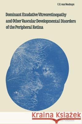
Dominant exudative vitreoretinopathy (DEVR) is an eye disease which has only recently received wider attention. In 1969 Criswick and Schepens used the designation "familial exudative vitreoretinopathy" to describe a syndrome they observed in six patients belonging to two families. The condition was characterized by several symptoms involving the vitreous and retina, e. g. "posterior vitreous detachment, organized vitreous membranes, heterotopia of the macula, retinal neovascularizations, subretinal and intraretinal exudates, and localized retinal detachment". The clinical features impressed the authors as strongly reminiscent of retrolental fibroplasia, but none of the patients had a record of premature birth or postnatal oxygen administration. In 1971 Cow and Oliver described the same syndrome in several members of one family. They considered their findings to be compatible with auto- somal dominant transmission. Canny and Oliver (I976) were the first to de- monstrate the fluorescein-angiographic changes of DEVR in four members of the abovementioned family. The most striking finding was "abrupt cessation of the capillary network in a scaloped edge near the equator". Fluorescein was seen to leak from the retinal vessels localized in this marginal zone, and in some eyes from massive fibrovascular lesions as well. Similar fluorescein- angiographic changes have been described in recent years in other reports on families with DEVR (Nijhuis et aI. , 1979; Slusher and Hutton, 1979; Dudgeon, 1979; Ober et a1. , 1980; Laqua, 1980). In 1979 I commenced a clinical study of this still little-known condition at the Nijmegen University Institute of Ophthalmology (The Netherlands).

Kategorie:
Nauka, Medycyna
Kategorie BISAC:
Medical > Okulistyka
Wydawca:
Springer
Seria wydawnicza:
Monographs in Ophthalmology
Język:
Angielski
ISBN-13:
9789400980235
Rok wydania:
2011
Wydanie:
Softcover Repri
Numer serii:
000162847
Ilość stron:
430
Waga:
0.66 kg
Wymiary:
23.5 x 15.5
Oprawa:
Miękka
Wolumenów:
01

I: The normal vasculature of the peripheral retina.- 1. Development and anatomy of the normal vasculature of the peripheral retina.- 1.1 Definitions of anatomical boundaries.- 1.2 Development and regression of the hyaloid vessels and the primary vitreous.- 1.3 Development of the choroid of the peripheral fundus.- 1.4 Development of the retinal vasculature.- 1.5 Anatomy of the vasculature of the peripheral retina.- 1.6 Structure of the retinal vessels and the vitreoretinal juncture of the peripheral fundus.- 2. Clinical examination of peripheral retinal blood vessels and circulation.- 2.1 Indirect ophthalmoscopy.- 2.2 Three-mirror contact lens examination.- 2.2.a The biomicroscopic features of the normal vasculature of the peripheral retina.- 2.3 Photography and fluorescein angiography of the peripheral fundus.- 2.3.a The fluorescein angiogram of the normal peripheral fundus.- II: Disturbed development of the peripheral retinal vessels.- 3. Clinical studies of families with dominant exudative vitreoretinopathy.- 3.1 Materials and methods.- 3.2 The A family.- 3.3 The B family.- 3.4 The C family.- 3.5 The D family.- 3.6 The E family.- 3.7 The F family.- 3.8 The G family.- 3.9 The H family.- 3.10 The I family.- 4. Results and discussion of the family studies.- 4.1 Numbers of affected and probably affected persons in the various families.- 4.2 Age and sex distribution of the affected and probably affected persons.- 4.3 Visual acuity.- 4.4 Refraction.- 4.5 Amblyopia.- 4.6 Nystagmus.- 4.7 Eye position.- 4.8 Intraocular pressure.- 4.9 Anterior segment.- 4.10 Vitreous.- 4.11 Fundus.- 4.11.a Biomicroscopic changes.- 4.11.b Fluorescein-angiographic changes.- 4.12 Visual fields.- 4.13 Ultrasonography.- 4.14 Electroretinography and electro-oculography.- 4.15 Dark adaptation.- 4.16 Colour vision.- 4.17 Histology.- 4.18 General physical examination.- 4.19 Stages and course.- 5. Genetic aspects and pathogenesis of dominant exudative vitreoretinopathy.- Genetic aspects.- 5.1 Mode of transmission.- 5.2 Gene penetrance.- 5.3 Gene expression.- 5.3.a Genetic factors.- 5.3.b Influence of the “environment”.- 5.4 Incidence and distribution of the gene.- 5.4.a Ethnic and geographical distribution.- 5.5 Genetic counselling.- Pathogenesis.- 5.6 DEVR as a vascular developmental disorder.- 5.7 Deformation of the foetal network of retinal vessels as direct consequence of disturbed vascular development. A hypothesis.- 5.8 Objections to qualification of DEVR as “vitreoretinal dystrophy”.- 6. Prevention and treatment of complications of dominant exudative vitreoretinopathy.- 6.1 Early diagnosis and selection of high-risk patients.- 6.2 Treatment.- 7. Congenital retinal fold (ablatio falciformis congenita).- 7.1 The clinical features of congenital retinal fold.- 7.2 Conditions which may be associated with congenital retinal fold.- 7.3 Congenital retinal fold as a manifestation of DEVR.- 7.4 Congenital retinal fold and microcephaly.- 7.5 Pathogenesis of congenital retinal fold.- 7.5.a Early versus late hypothesis.- 7.5.b Additional pathogenetic mechanisms.- 7.5.c Unilateral congenital retinal fold of obscure pathogenesis.- 8. Retrolental fibroplasia.- 8.1 Clinical symptoms.- 8.1.a Active retrolental fibroplasia.- 8.1.b Cicatricial retrolental fibroplasia.- 8.2 Histology.- 8.3 The influence of oxygen on the development of the immature retinal vasculature.- 8.4 Treatment.- 8.5 Differential diagnosis.- 8.5.a “Retrolental fibroplasia” without neonatal oxygen administration or prematurity.- 8.6 Consequences of oxygen administration to premature neonates with the genotype of DEVR.- 9. Affections with congenital or neonatal vitreous organizations and retinal detachment.- 9.1 Incontinentia pigmenti (Bloch-Sulzberger syndrome).- 9.1.a Clinical ocular symptoms.- 9.1.b Genetic aspects.- 9.1.c Histology and pathogenesis.- 9.1.d Differential diagnosis.- 9.1.e Treatment.- 9.2 Norrie’s disease.- 9.2.a Clinical symptoms.- 9.2.b Histology.- 9.2.c Pathogenesis.- 9.2.d Differential diagnosis.- 9.3 Reese-Blodi-Straatsma syndrome and trisomy 13.- 9.3.a Histology.- 9.3.b Pathogenesis.- 9.3.c Differential diagnosis.- 9.4 Congenital encephalo-ophthalmic dysplasia.- 9.4.a Clinical symptoms.- 9.5 Proliferative retinopathy in anencephalia.- 9.5.a Pathogenesis.- 9.6 Familial unilateral proliferative retinopathy.- 10. Observations on Wagner’s syndrome, sex-linked juvenile retinoschisis and juvenile rhegmatogenous retinal detachment.- 10.1 Wagner’s syndrome.- 10.1.a Clinical symptoms.- 10.1.b Histology.- 10.1.c General manifestations: clefting syndromes.- 10.1.d Fluorescein-angiographic study of the circulation of the equatorial retina and choroid.- 10.1.e Pathogenesis of the aberrant configuration of retinal vessels in the posterior pole and ectopia of the macula.- 10.2 Sex-linked juvenile retinoschisis.- 10.2.a Clinical symptoms.- 10.2.b Vascular changes.- 10.2.C Histology and pathogenesis.- 10.3 Juvenile rhegmatogenous retinal detachment.- 10.3.a Personal observations.- 11. Congenital bilateral arterial anastomosis between the choroid and the peripheral retina.- 12. Coats’ disease and retinal angiomatosis.- 12.1 Coats’disease.- 12.1.a Clinical symptoms.- 12.1.b Histology.- 12.1.c Pathogenesis.- 12.1.d Differential diagnosis.- 12.1.e Treatment.- 12.2 Retinal angiomatosis.- 12.2.a Clinical symptoms.- 12.2.b Histology and pathogenesis.- 12.2.C Differential diagnosis.- 12.2.d Treatment.- 12.3 Sturge-Weber’s syndrome.- III: Additional differential diagnoses from dominant exudative vitreoretinopathy.- 13. Congenital conditions not associated with retinal vascular developmental disorders.- 13.1 Persistent hyperplastic primary vitreous.- 13.1.a Clinical symptoms.- 13.1.b Histology.- 13.1.C Differential diagnosis.- 13.1.d Posterior PHPV and congenital retinal fold.- 13.2 Congenital toxoplasmosis.- 13.3 Retinoblastoma.- 14. Acquired conditions.- 14.1 Eales’disease.- 14.1.a Clinical symptoms.- 14.1.b Differential diagnosis from DEVR.- 14.2 Sickle cell retinopathy.- 14.2.a Differential diagnosis from DEVR.- 14.3 Pars planitis.- 14.3.a Clinical symptoms.- 14.3.b Differential diagnosis from DEVR.- 14.4 Toxocariasis.- 14.4.a Clinical symptoms.- 14.4.b Differential diagnosis.- 14.5 Myopia.- 14.5.a Perfusion disorders of the peripheral retina in myopia.- 14.5.b Ectopia of the macula in high myopia.- 14.6 Ectopia of the macula due to a pucker of the peripheral retina.- Summary.- Samenvatting.- References.- Curriculum vitae.


Udostępnij

Facebook - konto krainaksiazek.pl


Opinie o Krainaksiazek.pl na Opineo.pl

Partner Mybenefit

Krainaksiazek.pl w programie rzetelna firma Krainaksiaze.pl - płatności przez paypal

Czytaj nas na:

Facebook - krainaksiazek.pl
  • książki na zamówienie
  • granty
  • książka na prezent
  • kontakt
  • pomoc
  • opinie
  • regulamin
  • polityka prywatności

Zobacz:

  • Księgarnia czeska

  • Wydawnictwo Książkowe Klimaty

1997-2025 DolnySlask.com Agencja Internetowa

© 1997-2022 krainaksiazek.pl
     
KONTAKT | REGULAMIN | POLITYKA PRYWATNOŚCI | USTAWIENIA PRYWATNOŚCI
Zobacz: Księgarnia Czeska | Wydawnictwo Książkowe Klimaty | Mapa strony | Lista autorów
KrainaKsiazek.PL - Księgarnia Internetowa
Polityka prywatnosci - link
Krainaksiazek.pl - płatnośc Przelewy24
Przechowalnia Przechowalnia