Diatom Microscopy » książka


(netto: 922,32 VAT: 5%)
Najniższa cena z 30 dni: 962,48
ok. 30 dni roboczych.
Darmowa dostawa!
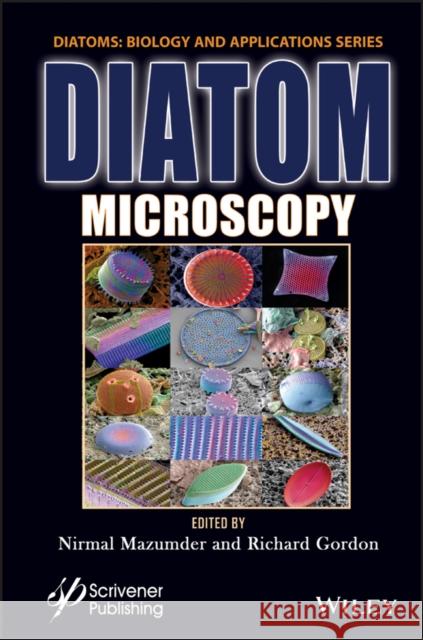
Preface xi1 Investigation of Diatoms with Optical Microscopy 1Shih-Ting Lin, Ming-Xin Lee and Guan-Yu Zhuo1.1 Introduction 21.2 Light Microscopy 41.2.1 Phase Contrast Microscopy 41.2.2 Differential Interference Contrast (DIC) Microscopy 61.2.3 Darkfield Microscopy 91.3 Fluorescence Microscopy 101.4 Confocal Laser Scanning Microscopy 121.5 Multiphoton Microscopy 191.6 Super-Resolution Optical Microscopy 221.7 Conclusion 25Acknowledgement 25References 252 Nanobioscience Studies of Living Diatoms Using Unique Optical Microscopy Systems 33Kazuo UmemuraAbbreviations 332.1 Trajectory Analysis of Gliding Among Individual Diatom Cells Using Microchamber Systems 352.2 Direct Observation of Floating Phenomena of Individual Diatoms Using a "Tumbled" Microscope System 402.3 Three-Dimensional Physical Imaging of Living Diatom Cells Using a Holographic Microscope System 44Acknowledgements 47References 473 Recent Insights Into the Ultrastructure of Diatoms Using Scanning and Transmission Electron-Microscopy 57Dharshini Gopal, Shweta Chakrabarti, Dataram Venkata Srikar Keshav, Richard Gordon and Nirmal Mazumder3.1 Introduction 583.2 Scanning Electron Microscopy (SEM) of Diatoms 583.3 Transmission Electron Microscopy (TEM) of Diatoms 653.3.1 Limitations 693.4 Conclusion 73References 734 Atomic Force Microscopy Study of Diatoms 81Ishita Chakraborty, Shweta Chakrabarti, Vishwanath Managuli and Nirmal Mazumder4.1 Introduction 824.2 Types of AFM Modes 864.3 Sample Preparation and Methods 884.4 Study of Diatom Ultrastructure Under AFM 884.5 Conclusion 102Glossary 104Acknowledgement 104References 1055 Refractive Index Tomography for Diatom Analysis 111Juan M. Soto, José A. Rodrigo and Tatiana Alieva5.1 Introduction 1125.2 Fundamentals of PC-ODT 1135.3 Experimental Setup for PC-ODT 1175.4 Diatom RI Reconstructions with Bright-Field Illumination 1205.5 Illumination Impact on PC-ODT Performance 1265.6 Concluding Remarks 131Acknowledgement 134References 1346 Luminescent Diatom Frustules: A Review on the Key Research Applications 139Jayur Tisso, Shruthi Shetty, Nirmal Mazumder, Ankur Gogoi and Gazi A. Ahmed6.1 Introduction 1406.2 Key Research Applications of Luminescence Properties of Diatom Frustules 1416.2.1 Novel Nanophotonic and Optoelectronic Applications of Luminescent Diatom Frustules 1426.2.2 Applications of Diatom Luminescence in Sensing 1456.2.3 Biomedical Applications of Diatom Luminescence 1496.2.4 Other Studies on Diatom Luminescence 1516.3 Future Perspectives 1676.4 Conclusion 168Acknowledgement 168References 1687 Micro to Nano Ornateness of Diatoms from Geographically Distant Origins of the Globe 179Mohd Jahir Khan, Daniel Mathys and Vandana Vinayak7.1 Introduction 1807.2 Materials and Methods 1837.2.1 Diatom Samples and Microscopy 1837.2.1.1 By Michael J. Stringer 1837.2.1.2 Diatom Oamaru Slides by Diane Winter 1847.2.1.3 By Daniel Mathys 1847.2.1.4 Diatom Sampling, Slide Preparation and Imaging from Himalayas, Plains and Arabian Sea, India 1857.3 Diatoms from Different Geographical Origins of the World 1857.3.1 Oamaru Diatoms 1857.3.2 Diatom Images Gifted by Michael J. Stringer 1867.3.3 Diatoms from Natural History Museum Basel, Switzerland a Piece of Art by Daniel Mathys 1907.3.4 Diatoms from India 1937.4 Conclusion 2167.5 Acknowledgements 216References 2168 Types of X-Ray Techniques for Diatom Research 221Mridula Sunder, Neha Acharya, Smitha Nayak, Richard Gordon and Nirmal Mazumder8.1 Introduction 2218.2 Applications 2228.2.1 Synchrotron Radiation-Based X-Ray Techniques 2228.2.2 X-Ray Computed Tomography 2248.2.3 X-Ray Fluorescence-Based Techniques 2268.2.4 X-Ray Microanalysis 2278.2.5 X-Ray Absorption-Based Techniques 2288.2.6 X-Ray Diffraction 2298.2.7 Other X-Ray-Based Techniques 2308.3 Conclusions 233Glossary 233References 2349 Diatom Assisted SERS 237Rajib Biswas and Sankar Biswas9.1 Introduction 2379.2 Diatom 2399.2.1 Basic Overview 2399.2.2 Physiological Characteristics 2399.2.3 Optical and Relevant Properties 2409.3 Raman Scattering 2419.3.1 Basics 2419.3.2 Surface Enhanced Raman Scattering 2429.3.3 Optoelectronic Investigations 2429.4 SERS Through Diatom: Fundamentals and Application Overview 2439.5 Conclusion and Future Outlook 245References 24610 Diatoms as Sensors and Their Applications 251Priyasha De and Nirmal Mazumder10.1 Introduction 25110.2 Diatoms as Biosensors 25510.2.1 Electrochemical Sensors 26010.2.2 Plasmonic Sensors 26110.2.3 Immunoassay Sensors 26410.2.4 Optical and Optofluidic Sensors 26810.2.5 Biochemical Sensors 27110.2.6 FRET-Based Sensors 27310.2.7 Microfluidics-Based Sensors 27410.3 Conclusion 276Acknowledgments 276References 27711 Diatom Frustules: A Transducer Platform for Optical Detection of Molecules 283Viji S., Ponpandian N. and Viswanathan C.11.1 Introduction 28411.2 Optical Properties of Diatom Frustules 28511.2.1 Diatom as a Photoluminescent Materials 28511.2.2 Diatom as a Photonic Crystal 28711.2.3 Diatoms as a SERS Substrate 28811.3 Methods Involved in Thin Film Deposition of Diatom Frustules 29011.4 Diatom as an Optical Transducer for Biosensors 29411.5 Diatom as an Optical Transducer for Gas/Chemical Sensors 29711.6 Conclusion 300References 30112 Effects of Light on Physico-Chemical Properties of Diatoms 307Janardan Sen, Priyal Dhawan, Priyasha De and Nirmal Mazumder12.1 Introduction 30812.2 Effect of Light on Diatom Function and Morphology 31012.2.1 Effect of Light Intensity on Diatom Morphology 31012.2.2 Effect of Light Intensity on Diatom Growth 31312.2.3 Effect of Light Intensity on Photosynthesis in Diatoms 31812.2.4 Effect of Wavelength of Light on Diatom Pigment System 32112.2.5 Effect of Light Intensity on the Physiology of Diatoms 32712.3 Conclusion 330Acknowledgment 330References 330Index 335
Nirmal Mazumder obtained his PhD degree in 2013 from National Yang-Ming University, Taiwan. He has two years of post-doctoral research experience. In 2016, he joined the Department of Biophysics, Manipal School of Life Sciences, Manipal Academy of Higher Education, Manipal, India as an assistant professor. His research interests relate to the development of Stokes-Mueller-based light microscopy for tissue characterization and deep learning. He also investigates the photonics properties of diatoms as well as frustules for bio-photonics applications. He has published several peer-reviewed journal articles and book chapters.Richard Gordon's involvement with diatoms goes back to 1970 with his capillarity model for their gliding motility, published in the Proceedings of the National Academy of Sciences of the United States of America. He later worked on a diffusion-limited aggregation model for diatom morphogenesis, which led to the first paper ever published on diatom nanotechnology in 1988. He organized the first workshop on diatom nanotech in 2003.
1997-2026 DolnySlask.com Agencja Internetowa
KrainaKsiazek.PL - Księgarnia Internetowa


