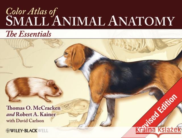
Color Atlas of Small Animal Anatomy: The Essentials » książka


Color Atlas of Small Animal Anatomy: The Essentials
ISBN-13: 9780813816081 / Angielski / Miękka / 2009 / 160 str.
Color Atlas of Small Animal Anatomy: The Essentials
ISBN-13: 9780813816081 / Angielski / Miękka / 2009 / 160 str.
(netto: 315,02 VAT: 5%)
Najniższa cena z 30 dni: 328,73
ok. 22 dni roboczych.
Darmowa dostawa!
Extraordinary accuracy and original artwork are just two features readers will find in this new resource, providing a basic foundation in small animal anatomy. Its unique organization includes the anatomy of all organ systems in the dog, cat, rabbit, rat and guinea pig.
"This book will be an invaluable resource for veterinary students, teachers, and practitioners alike as it manages to present information in a manner that is easy to understand by almost any reader". (Mammalia, 2010)
"This atlas fills the gap between... highly detailed references and oversimplified anatomic descriptions. The authors have achieved their objectives by providing accurate descriptions of the most pertinent anatomy while avoiding excessive detail... it manages to present information in a fashion that is easy to understand by almost any reader. Veterinary students will appreciate the comparative nature of the book, with the range of species presented. Professionals with more advanced knowledge will find the atlas useful in explaining anatomy to laypersons, clients, and students." – Doody′s Reviews, June 2009
The Color Atlas of Small Animal Anatomy: The Essentials beautifully depicts the topographic anatomy of organ systems in dogs, cats, rabbits, rats, and guinea pigs.... The atlas is an invaluable source of accurate basic anatomic illustrations for veterinary medical students, practitioners, and educators and contains reasonable details to be of use for laboratory animal researchers. – Journal of the American Veterinary Medical Association
"I would recommend this book for any clinic or hospital and suggest that your technicians and staff will use it often." – Veterinary Information Network
Section 1.
The Dog.
Plate 1.1 Lateral view of the dog (Beagle).
Plate 1.2 Lateral view of the bitch (Retriever).
Plate 1.3 Body regions.
Plate 1.4 Skeleton.
Plate 1.5 Cutaneous muscles and major fasciae the dog.
Plate 1.6 Superficial muscles of the bitch.
Plate 1.7 Deep muscles of the dog.
Plate 1.8 Deep cervical muscles, major joints, and in situ viscera of the bitch.
Plate 1.9 Paraxial view of the third digit.
Plate 1.10 Palmar views of the major structures of the forepaw; plantar view of the major structures of the hidpaw.
Plate 1.11 Median section of the head, and dentition.
Plate 1.12 The eye and accessory ocular structures.
Plate 1.13 The nose.
Plate 1.14 The ear.
Plate 1.15 Mouth and tongue and esophagus.
Plate 1.16 Ventral view of the abdomen and its structures.
Plate 1.17 Large intestine, anus and anal sacs.
Plate 1.18 Body cavities and serous membranes.
Plate 1.19 Thoracic, abdominal and pelvic viscera related to the skeleton of the dog.
Plate 1.20 Thoracic, abdominal and pelvic viscera, and mammary glands of the bitch.
Plate 1.21 Hip joint.
Plate 1.22 Location of major endocrine organs.
Plate 1.23 Relations of the reproductive organs of the dog.
Plate 1.24 Relations of the reproductive organs of the bitch.
Plate 1.25 Major veins.
Plate 1.26 Major arteries.
Plate 1.27 Lymph nodes and vessels.
Plate 1.28 Central and somatic nervous system.
Plate 1.29 Autonomic nervous system.
Plate 1.30 Brain, dorsal, ventral and lateral views.
Section 2.
The Cat.
Plate 2.1 Lateral view of the male cat (Moggie–nonpedigree).
Plate 2.2 Lateral view of the female cat (Persian).
Plate 2.3 Endocrine organs and lymph nodes.
Plate 2.4 Skeleton.
Plate 2.5 Cutaneous muscles and major fasciae of the male.
Plate 2.6 Superficial muscles of the female.
Plate 2.7 Middle muscles and in situ viscera of the male.
Plate 2.8 Deep muscles and in situ viscera of the female.
Plate 2.9 Median section of the head, and dentition.
Plate 2.10 Oral cavity, tongue, pharynx and esophagus.
Plate 2.11 The external, middle, and inter ear.
Plate 2.12 The eye and accessory ocular structures.
Plate 2.13 Isolated stomach and intestines.
Plate 2.14 Large intestine, anus and anal sacs.
Plate 2.15 Superficial and deep structures of the paw (foot) lateral view.
Plate 2.16 Plantar views of the major structures of forepaw and hindpaw.
Plate 2.17 Thoracic, abdominal and pelvic viscera related to the skeleton of the male.
Plate 2.18 Thoracic, abdominal and pelvic viscera, related to the skeleton of the female.
Plate 2.19 Relations of the reproductive organs of the male.
Plate 2.20 Relations of the reproductive organs of the female.
Plate 2.21 Major veins.
Plate 2.22 Major arteries.
Plate 2.23 Central and peripheral nervous system.
Plate 2.24 Brain, dorsal, ventral and lateral views.
Section 3.
The Rabbit.
Plate 3.1 Lateral view.
Plate 3.2 Body regions.
Plate 3.3 Skeleton.
Plate 3.4 Endocrine organs and lymph nodes.
Plate 3.5 Superficial muscles of the male.
Plate 3.6 Deep muscles of the female.
Plate 3.7 Median section of the rabbit s head and dentition.
Plate 3.8 Oral cavity, tongue, pharynx and esophagus.
Plate 3.9 Thoracic, abdominal and pelvic viscera (in situ) of the male.
Plate 3.10 Thoracic, abdominal and pelvic viscera (in situ) of the female.
Plate 3.11 Relations of the reproductive organs of the male.
Plate 3.12 Relations of the reproductive organs of the female.
Plate 3.13 Central and peripheral nervous system.
Plate 3.14 Brain, dorsal, ventral, and lateral views.
Section 4.
The Rat.
Plate 4.1 Lateral view.
Plate 4.2 Skeleton of the rat.
Plate 4.3 Superficial muscles of the male.
Plate 4.4 Deep and middle muscles of the female.
Plate 4.5 Median section of the head and dentition.
Plate 4.6 Ventral view of abdominal structures (in situ) and diagram of digestive system.
Plate 4.7 Thoracic, abdominal and pelvic viscera related to the skeleton of the male.
Plate 4.8 Thoracic, abdominal and pelvic viscera, related to the skeleton of the female.
Plate 4.9 Relations of the reproductive organs of the male.
Plate 4.10 Relations of the reproductive organs of the female.
Plate 4.11 Spinal nerves.
Plate 4.12 Autonomic nerves.
Plate 4.13 Brain, dorsal, ventral, and lateral views.
Plate 4.14 Brian, sagittal section, and detail of midbrain.
Section 5.
The Guinea Pig.
Plate 5.1 Lateral view.
Plate 5.2 Skeleton.
Plate 5.3 Superficial muscles of the male.
Plate 5.4 Deep and middle muscles of the female.
Plate 5.5 Median section of the head and dentition.
Plate 5.6 Ventral view of abdominal structures (in situ) and diagram of digestive system.
Plate 5.7 Thoracic, abdominal and pelvic viscera related to the skeleton of the male.
Plate 5.8 Thoracic, abdominal and pelvic viscera, and mammary glands of the female.
Plate 5.9 Relations of the reproductive organs of the male.
Plate 5.10 Relations of the reproductive organs of the female.
Plate 5.11 Central and peripheral nervous system.
Plate 5.12 Brain dorsal, ventral, and lateral views
Thomas O. McCracken, MS, is Professor of Anatomy & Physiology at Robert Ross International University of Nursing (IUON) in Basseterre, St Kitts, West Indies; and Former Associate Professor of Anatomy at the College of Veterinary Medicine and Biomedical Sciences, Colorado State University.
Robert A. Kainer, DVM, MS, is Professor Emeritus of Anatomy at the College of Veterinary Medicine and Biomedical Sciences, Colorado State University, Fort Collins, Colorado.
This new resource provides a basic foundation in small animal anatomy for students of veterinary medicine, animal science, and veterinary technology. Extraordinary accuracy and beautiful original artwork make this a truly unique learning tool that includes the anatomy of all organ systems in the dog, cat, rabbit, rat, and guinea pig – all described in a consistent manner.
Learning features include: carefully selected labeling helps students learn and remember structures and relationships; male and female of species are depicted on facing pages so topographic anatomy can be compared; structures common to various animals are labeled several times, whereas unique structures are labeled on one or two species so students can make rapid distinctions of the structures peculiar to certain animals; and an introduction that provides readers with a background in nomenclature and anatomic orientation so they can benefit from the atlas even if they lack training in anatomy.
The Atlas depicts topographic relationships of major organs in a simple, yet technically accurate presentation that′s free from extraneous material so that those using the atlas can concentrate on the essential aspects of anatomy. It will be an invaluable resource for veterinary students, teachers and practitioners alike.
1997-2026 DolnySlask.com Agencja Internetowa
KrainaKsiazek.PL - Księgarnia Internetowa


