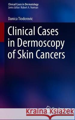
Clinical Cases in Dermoscopy of Skin Cancers » książka


Clinical Cases in Dermoscopy of Skin Cancers
ISBN-13: 9783030294465 / Angielski / Miękka / 2020 / 225 str.
Clinical Cases in Dermoscopy of Skin Cancers
ISBN-13: 9783030294465 / Angielski / Miękka / 2020 / 225 str.
(netto: 192,11 VAT: 5%)
Najniższa cena z 30 dni: 192,74
ok. 22 dni roboczych
Bez gwarancji dostawy przed świętami
Darmowa dostawa!
“The intended audience includes any medical practitioners who employ dermoscopy of the skin in their practice. ... This book would be a valuable addition to the field of dermoscopy … .” (Renata H. Mullen, Doody’s Book Reviews, March 27, 2020)
67-year-old man with a pigmented lesion on the left temporal region
Invasive lentigo maligna in a 72-year-old man
A 36-year-old man with growing pigmented lesion
A 46-year-old woman presented to the office asking for evaluation of pigmented lesion on her face
A 70-year-old farmer with pigmented lesion on the cheek
A changing pigmented lesion in a 41-year-old woman
A newly developing pigmented lesion in elderly patient – the importance of clinic pathologic correlation
Extrafacila lentigo maligna melanoma located on the back of a 65-year old man
Importance of full-body examination in melanoma detection in early detection of melanoma
Two superficial spreading melanomas at the same time in the same patient
Melanoma hidden amongst seborrheic keratosis
A 28-year-old man presented to the office asking for evaluation of a pigmented lesion on his pectoral region
A changing pigmented lesion on the left glueteal region in a 67-year-old man
A melanoma rising in congenital melanocytic nevus in a 58-year-old man
Melanocytic nevus-like melanoma in a 38-year old patient
A 35-year-old woman presented to the office asking for evaluation of pigmented lesion located on her left upper arm
Dysplastic nevus syndrome associated with melanoma in a 45-year-old patient
A 43-year-old patient with flat pigmented lesion located on the back
Small diameter melanoma in a 38-year old patient
A 28-year-old man presented to the office asking the treatment of pityriasis versicolor infection having a small diameter melanoma on the scalp at the same time
A peripherally distributed dots as a sign for melanoma detection in a 41-year-old patient
Blue nevus like melanoma
A 58-year-old woman presented to the office asking for evaluation of nodular lesion located on the right leg
A 92-year old patient with pigmented nodular lesion on the left temporal region
Nodular lesion located on the back in a 64-year old patient
Growing nodular lesion on the back of a 52-year old man
A 68-year-old patient with a changing congenital melanocytic nevus
A melanoma resembling basal cell carcinoma
An apigmented flat lesion on the abdomen
A misdiagnosed acral melanoma
Acral lentiginous melanoma presented as an interdigital erosion in a 38-year-old patient
A non-pigmented flat lesion located on the abdomen in a 62-year-old patient
A 68-year-old patient with flat apigmented lesion located on the back
Invisible basal cell carcinoma located on the right forearm in a 78-year-old patient
A 68-year-old man presented to the office asking for evaluation of erythematous plaque located on the right subscapular region
Two basal cell carcinomas resembling dermal nevi
A 66-year-old patient with a nodular apigmented lesion
Invisible basal cell carcinoma located on the face in a 49-year-old patient
An 81-year-old patient with two basal cell carcinomas on the face
Highly pigmented lesion on the back in a 46-year-old patient
A 57-year-old man with linear pigmented lesion located on his neck
A 63-year-old patient with a large pigmented lesion located on the back
A 72-year old patient with nodular pigmented lesion located on the right lateral side of the nose
Whitish plaque located on the nose in a 82-year-old patient
A 58-year-old patient with non-pigmented lesion located on the left cheek
A 49-year-old patient with pigmented flat lesion located on the nose
A 71-year-old patient with flat hypopigmented lesion located on left temporal region
A 69-year-old patient with non-pigmented flat leson located on the leg
Two apigmented slightly elevated lesions in a 42-year-old patient
A 65-year old patient with growing apigmented lesion located on the right cheek
A 68-year-old patient with newly developing apigmented lesion located on the right leg
A 72-year-old patient with non-pigmented bleeding leson on the nose
Danica Tiodorovic, PhD, MD, was born in Nis, Serbia on July 14, 1978. She enrolled at the medical faculty at the University of Nis in 1997. She graduated on March 1, 2004 with an average mark 9.95 and graduated with honors (mark 10 for the final exam in dermatovenerology). She enrolled at the postgraduate studies in 2004 with dermatovenerology being my major. She received scholarship from the Ministry of Science and Ecology which was active up to my employment at the Clinic of Dermatovenerology on May 20, 2005 in Nis. She received a master’s degree on May 3, 2007. The master’s degree thesis was, Liability estimate of dermoscopy as additional diagnostic method in clinical examination of small skin tumors“. She finished my specialization with excellent marks on June 1, 2010. In 2007 she was granted a doctor's thesis with the title "Correlation of global pattern, pigment distribution and color of melanocytic naevi determined by dermoscopy with a skin type and age". Her special field of interest is dermoscopy, the field which she specialized in and did research at the Department of Dermatology, with Prof. Giuseppe Argenziano and Prof. Iris Zalaudek in Naples, Rome, Reggio Emliia and Graz and Prof. Harald Kittler in Vienna. She has been invited to lecturer at the Congresses of European Academy of Dermatology, American Academy of Dermatology and World Congresses of Dermoscopy. She became a Representative of the Republic of Serbia in the International Dermoscopy Society (IDS) and Board member of International Dermoscopy Society (IDS). In 2015 she become Secretary General of Serbian Association of Dermatovenerology.
This book provides a practical guide to the clinical decision-making process used in the management of skin cancers with the use of dermoscopy. Clinical cases are examined to help the reader through the treatment of unusual skin cancers using best practice techniques. A variety of skin conditions are covered, including melanoma, basal cell carcinoma, squamous cell carcinoma, Bowen’s disease and actinic keratosis.
Clinical Cases in Dermoscopy of Skin Cancers highlights evidence-based best practice through its multidisciplinary approach and is an important addition to the literature to help trainees and practicing dermatologists or any healthcare professional who manages these patients.
1997-2025 DolnySlask.com Agencja Internetowa
KrainaKsiazek.PL - Księgarnia Internetowa


