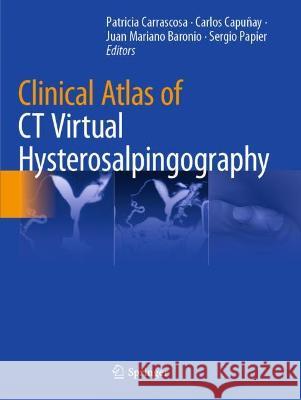
Clinical Atlas of CT Virtual Hysterosalpingography » książka


Clinical Atlas of CT Virtual Hysterosalpingography
ISBN-13: 9783030662097 / Angielski / Miękka / 2022
This book provides a comprehensive, practically applicable guide to the use of CT virtual hysterosalpingography for evaluating gynaecological pathology and infertility in women. It features detailed descriptions of normal and pathologic findings across the female reproductive system, including the cervix, uterine wall and cavity, and Fallopian tubes, and compares the findings with other imaging modalities such as ultrasound, X-ray hysterosalpingography and MRI. The interpretation of post-treatment findings and commonly encountered pitfalls are also covered in detail. Clinical Atlas of CT Virtual Hysterosalpingography compares the use of a variety of imaging modalities to assess female infertility, and is a valuable resource for medical professionals who encounter these patients in their clinical practice.
Computed Tomography Virtual Hysterosalpingography.- Normal Radiologic Anatomy of Female Reproductive System.- Case 1, Normal Anatomy.- Case 2, Normal Anatomy.- Case 3, Normal Anatomy.- Case 4, Normal Anatomy.- Case 5, Normal Anatomy.- Case 6, Normal Anatomy.- Case 7, Normal Anatomy.- Case 8, Normal Anatomy.- Case 9, Retroversio-retroflexio Uterus.- Case 10, Anteversio-retroflexio Uterus.- Case 11, Retroversio-retroflexio Uterus.- Case 12, Intrauterine Device.- Case 13, Cervical Balloon Cannula.- Case 14, Normal Anatomy.- Case 15, Normal Anatomy.- Case 16, Normal Anatomy.- Case 17, Cornual Lucencies.- Evaluation of the Cervix.- Case 18, Focal Cervical Stenosis.- Case 19, Focal Cervical Stenosis.- Case 20, Focal Cervical Stenosis.- Case 21, Cervical Stenosis in Arcuate Uterus.- Case 22, Uterine Cervix Stenosis.- Case 23, Uterine Cervix Stenosis.- Case 24, Uterine Cervix Stenosis.- Case 25, Focal Cervical Synechiae.- Case 26, Cervical Synechiae.- Case 27, Cervical Synechiae.- Case 28, Cervical Synechiae.- Case 29, Cervical Synechiae.- Case 30, Cervical Synechiae.- Case 31, Cervical Synechiae.- Case 32, Hypertrophic Cervical Folds.- Case 33, Cervical Polyp.- Case 34, Cervical Polyp.- Case 35, Cervical Polyp .- Case 36, Cervical Polyp.- Case 37, Cervical Polyp.- Case 38, Cervical Polyp.- Case 39, Cervical Gland Dilation.- Case 40, Cervical Gland Diverticula.- Case 41, Cervical Gland Diverticula.- Case 42, Cervical Gland Diverticula.- Pathology of the Uterine Cavity.- Case 43, Focal Intrauterine Synechiae.- Case 44, Uterine Synechiae.- Case 45, Uterine Synechiae.- Case 46, Uterine Synechiae.- Case 47, Diffuse Uterine Synechiae.- Case 48, Diffuse Uterine Synechiae.- Case 49, Intrauterine Synechiae.- Case 50, Diffuse Uterine Synechiae.- Case 51, Diffuse Uterine Synechiae.- Case 52, Diffuse Uterine Synechiae.- Case 53, Uterine Synechiae and Cervical Stenosis.- Case 54, Uterine Synechiae in a Bicomuate Uterus.- Case 55, Endometrial Hyperplasia.- Case 56, Endometrial Hyperplastic Fold.- Case 57, Endometrial Polyp.- Case 58, Endometrial Polyp.- Case 59, Endometrial Polyp.- Case 60, Endometrial Polyp.- Case 61, Endometrial Polyp.- Case 62, Endometrial Polyp.- Case 63, Endometrial Polyp.- Case 64, Endometrial Polyp.- Case 65, Endometrial Polyp.- Case 66, Endometrial Polyp.- Case 67, Pedunculated Endometrial Polyp.- Case 68, Endometrial Polyp.- Case 69, Endometrial Polyp in a Bicornuate Uterus.- Case 70, Endometrial Polyp.- Case 71, Endometrial Polyp.- Case 72, Endometrial Polyp.- Case 73, Endometrial Polyp.- Case 74, Large Pedunculated Endometrial Polyp.- Case 75, Endometrial Polyps.- Case 76, Multiple Sessile Endometrial Polyps.- Case 77, Multiple Polyps.- Case 78, Multiple Endometrial Polyps.- Case 79, Multiple Endometrial Polyps.- Case 80, Submucosal Myoma.- Case 81, Submucosal Myoma.- Case 82, Submucosal Myoma.- Case 83, Submucosal Myoma.- Case 84, Submucosal Myoma.- Case 85, Submucosal Myomas.- Case 86, Submucosal Myoma.- Case 87, Submucosal Myoma.- Case 88, Submucosal Myoma.- Case 89, Submucosal Myoma.- Case 90, Submucosal Myoma.- Case 91, Submucosal Leiomyoma: Tubal Obstruction.- Case 92, Submucosal Myomas.- Pathology of the Uterine Wall.- Case 93, Intramural Myoma.- Case 94, Hybrid Myoma.- Case 95, Hybrid Myoma.- Case 96, Hybrid Myoma.- Case 97, Subserosal Myoma with Bilateral Hydrosalpinx.- Case 98, Subserosal Myoma.- Case 99, Subserosal Myoma.- Case 100, Focal Adenomyosis.- Case 101, Adenomyosis.- Case 102, Focal Adenomyosis.- Case 103, Adenomyosis.- Case 104, Fundal Adenomyosis.- Case 105, Focal Fundal Adenomyosis.- Case 106, Fundal Adenomyosis.- Case 107, Diffuse Adenomyosis.- Case 108, Intravasation of Contrast Media.- Case 109, Intravasation of Contrast Media.- Case 110, Intravasation of Contrast Media.- Congenital Uterine Anomalies.- Case 111, Unicornuate Uterus.- Case 112, Unicornuate Uterus.- Case 113, Unicornuate Uterus.- Case 114, Unicornuate Uterus.- Case 115, Unicornuate Uterus.- Case 116, Bicornuate Uterus (Unicollis).- Case 117, Bicornuate Uterus.- Case 118, Bicornuate Uterus.- Case 119, Bicornuate Uterus.- Case 120, Didelphys Uterus.- Case 121, Uterus Didelphys with Partial Vaginal Septation.- Case 122, Arcuate Uterus.- Case 123, Arcuate Uterus.- Case 124, Arcuate Uterus.- Case 125, Partial Septate Uterus.- Case 126, Partial Septate Uterus.- Case 127, Partial Septate Uterus.- Case 128, Partial Septate Uterus.- Case 129, Complete Septate Uterus with Partial Communication.- Case 130, Complete Septate Uterus.- Case 131, T-shaped Uterus.- Case 132, T-shape Uterus with Right Fallopian Tube Dilatation.- Pathology of the Fallopian Tubes.- Case 133, Left Fallopian Tube Dilatation with Intratubal Synechiae.- Case 134, Left Tubal Dilatation and Focal Adenomyosis.- Case 135, Mild Left Hydrosalpinx.- Case 136, Right Tube Hydrosalpinx.- Case 137, Left Tubal Occlusion and Right Hydrosalpinx.- Case 138, Right Tube Hydrosalpinx.- Case 139, Right Hydrosalpinx.- Case 140, Mild Right Ampulla Dilatation and Focal Adenomyosis.- Case 141, Left Hydrosalpinx.- Case 142, Right Hydrosalpinx with Tubal Synechiae.- Case 143, Mild Right Hydrosalpinx.- Case 144, Bilateral Tubal Synechiae.- Case 145, Hemaosalpinx, Adenomyosis, and Endometriosis.- Case 146, Bilateral Hydrosalpinx.- Case 147, Bilateral Hydrosalpinx.- Case 148, Bilateral Hydrosalpinx.- Case 149, Bilateral Hydrosalpinx.- Case 150, Tubal Obstruction, Uterine Synechiae, Endometrial and Cervical Polyps.- Case 151, Right Fallopian Tube Dilatation and Left Hydrosalpinx with Tubal Polyp.- Case 152, Left Tubal Polyp, Right Tubal Synechiae and Endometrial Polyp.- Case 153, Left Salpingitis Ishmica Nodosum.- Morphological Postsurgical Changes.- Case 154, Myoma Resection.- Case 155, Asherman Syndrome Secondary to Myoma Resection.- Case 156, Late Complication of Myomectomy: Paracervical Sacculation After Myomectomy.- Case 157, Uterine Septoplasty.- Case 158, Uterine Septoplasty.- Case 159, Uterine Septoplasty.- Case 160, Uterine Septoplasty.- Case 161, Bicornuate Uterus.- Case 162, Sequel of Hyteroscopic Synechiae Resection.- Case 163, Sequel of Surgical Uterine Synechiae Resection.- Case 164, Sequel of Surgical Uterine Synechiae Resection.- Case 165, Casarean Section Scar.- Case 166, Casarean Section Isthmocele.- Case 167, Casarean Section Isthmocele.- Case 168, Casarean Section Isthmocele.- Case 169, Casarean Section Isthmocele.- Case 170, Vesico-uterine Fistula Post-casarean Section.- Case 171, Right Salpingectomy.- Pitfalls and Incidental Findings.- Case 172, Pitfalls: Myometrial Folds.- Case 173, Pitfalls: Myometrial Folds.- Case 174, Pitfalls: Secretions Within the Endometrial Cavity.- Case 175, Pitfalls: Intrauterine Blood Remnants.- Case 176, Pitfalls: Intrauterine Blood Clot.- Case 177, Small Air Bubble.- Case 178, Air Bubbles.- Case 179, Air Bubbles.- Case 180, Air Bubbles.- Case 181, Large Air Bubble.- Case 182, Ovarian Cyst.- Case 183, Ovarian Teratoma.- Case 184, Ovarian Teratoma.- Case 185, Left Ovarian Teratoma and Hydrosalpinx.
Patricia Carrascosa, MD, PhD, FSCCT, FACC, MSCCT is Medical Director and Head of Research Department of Diagnóstico Maipú-DASA (one of the main imaging centers in Argentina). She is currently assistant professor of Buenos Aires University in Argentina. She obtained her PhD in Virtual Colonoscopy in 2007. She has been working in research in 3D special studies (virtual studies, angiographies) and since 1999 she has focused her main activity in the cardiovascular field in CT and MRI. She is boarded in Cardiovascular Computed Tomography and she is Fellowship and Master of the Society of Cardiovascular Computed Tomography as well as Co-President of the Latinamerican Committee of the SCCT. Author of many scientific publications, she also participates in international trials. Dr Carrascosa has over 100 manuscripts published in national and international journals. She has written 5 books: La enfermedad Coronaria en Tomografía Computarizada, Colonoscopía Virtual, CT Virtual Hysterosalpingography, Dual Energy CT in Cardiovascular and Clinical Atlas of Cardiac and Aortic CT and MRI. She is also author of national and international books chapters about different subjects in the radiology field.
Carlos Capuñay, MD, is currently Head of CT-MRI Department and Sub-head of Research Department of Diagnóstico Maipú-DASA in Buenos Aires, Argentina. He completed his medical studies in 1995 at Universidad del Salvador in Buenos Aires and his medical residency in Diagnostic Imaging in 1998 at Diagnóstico Maipú. He has been working in research on CT virtual studies and cardiovascular examinations since 2000. He is certified and three-times recertified in Radiology by the Argentine Council of Evaluation in Diagnostic Imaging (CONAEDI) and he is board certified in Cardiovascular Computed Tomography. He has over 90 manuscripts published in national and international peer-reviewed journals and he has written 4 books: La enfermedad Coronaria en Tomografía Computarizada, Colonoscopía Virtual, CT Virtual Hysterosalpingography and Clinical Atlas of Cardiac and Aortic CT and MRI. He is also author of national and international books chapters about different subjects in the radiology field.Juan Mariano Baronio, MD, has completed his medical studies in Universidad del Salvador in Buenos Aires, Argentina. He is specialist in gynaecology and obstetrics and in gynaecological and reproductive endocrinology. He has a Master's Degree in Reproductive Medicine and he is also a certified laparoscopy and hysteroscopy surgeon. Dr. Baronio has participated in numerous congresses, conferences as well as national and international courses of the specialty. He is author and co-author in several scientific works and has written the book: CT Virtual Hysterosalpingography.
Sergio Papier, MD, is currently Medical Director of CEGYR. He has completed his medical studies in Buenos Aires University in Argentina. He is specialist in gynaecology and reproductive medicine. Former President of the Argentine Society of Reproductive Medicine (SAMER) - President of the Latin American Association of Reproductive Medicine (ALMER).
This book provides a comprehensive, practically applicable guide to the use of CT virtual hysterosalpingography for evaluating gynaecological pathology and infertility in women. It features detailed descriptions of normal and pathologic findings across the female reproductive system, including the cervix, uterine wall and cavity, and Fallopian tubes, and compares the findings with other imaging modalities such as ultrasound, X-ray hysterosalpingography and MRI. The interpretation of post-treatment findings and commonly encountered pitfalls are also covered in detail.
Clinical Atlas of CT Virtual Hysterosalpingography compares the use of a variety of imaging modalities to assess female infertility, and is a valuable resource for medical professionals who encounter these patients in their clinical practice.
1997-2025 DolnySlask.com Agencja Internetowa
KrainaKsiazek.PL - Księgarnia Internetowa


