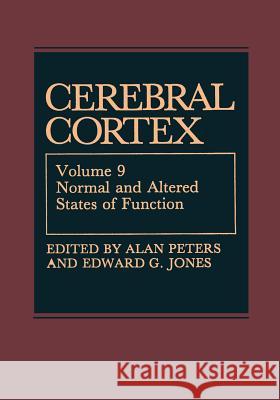
Cerebral Cortex: Normal and Altered States of Function » książka


Cerebral Cortex: Normal and Altered States of Function
ISBN-13: 9781461566243 / Angielski / Miękka / 2012 / 535 str.
Cerebral Cortex: Normal and Altered States of Function
ISBN-13: 9781461566243 / Angielski / Miękka / 2012 / 535 str.
(netto: 382,46 VAT: 5%)
Najniższa cena z 30 dni: 385,52
ok. 22 dni roboczych.
Darmowa dostawa!
This volume of the series on "Cerebral Cortex" deals with a variety of topics that need to be considered in our overall understanding of the functions of the cerebral hemispheres. Chapters in the first part of this volume deal with normal functions that were not covered in earlier volumes, while chapters in the latter part deal with the functioning of the cortex in various altered states. The first chapter is by Eberhard Fetz, Keisuke Toyama, and Wade Smith, and it considers the interactions that can be demonstrated to exist between cortical neurons by using the technique of cross-correlation. The second chapter is by Brent Vogt who examines the connections and functions of layer I of the cerebral cortex, a layer that has been largely ignored in the past, and he proposes that this layer probably plays an important role in learning and memory acquisi tion. This is followed by a chapter in which Oswald Steward presents a review of what is currently known about synaptic replacement following denervation of cortical neurons, and especially those in the hippocampus."
1 Synaptic Interactions between Cortical Neurons.- 1. Introduction.- 1.1. Measures of Synaptic Interactions.- 1.2. Effects of Synaptic Connections.- 2. Visual Cortex.- 2.1. Application of the Cross-Correlation Technique in the Visual System.- 2.2. Geniculocortical Interaction.- 2.3. Corticogeniculate Interaction.- 2.4. Intra- and Intercolumnar Interaction in Single and Adjacent Functional Columns of the Visual Cortex.- 2.5. Transcolumnar Interaction between Distant Columns.- 2.6. Synaptic Interaction Demonstrated by STA.- 2.7. Functional Conclusions.- 3. Auditory Cortex.- 4. Somatosensory Cortex.- 4.1. Cross-Correlation Studies.- 4.2. STA Studies.- 5. Motor Cortex.- 5.1. Cross-Correlation Studies.- 5.2. STA Studies.- 6. Association Cortex.- 7. Hippocampus.- 7.1. Cross-Correlation Studies.- 7.2. STA Studies.- 8. Summary and Conclusions.- 8.1. Common Features of Synaptic Interactions.- 8.2. Future Directions.- 9. References.- 2 The Role of Layer I in Cortical Function.- 1. Introduction.- 2. Behavioral Role of Layer I.- 3. Layer I in Sensory Cortices.- 3.1. Electrophysiology of Layer II Neurons.- 3.2. Afferent Connections and Physiology.- 4. Architecture of Layer I.- 4.1. Subdivisions.- 4.2. Neuronal Composition.- 4.3. Compartmentation of Apical Dendrites.- 5. The Proximal and Distal GABAergic Systems.- 6. Compartmentation of Afferent Connections.- 6.1. Thalamic and Subicular Projections.- 6.2. Serotoninergic Projections to Layer I.- 7. Passive and Active Interactions between Distal and Proximal Dendritic Compartments.- 8. Cholinergic Projections: Organization and Role in Event Holding.- 9. Noradrenergic Projections to Layer I and Memory Consolidation.- 10. What Is the Role of Layer I in Cortical Function?.- 11. References.- 3 Synapse Replacement on Cortical Neurons following Denervation.- 1. Introduction.- 2. The Process of Reinnervation in the Dentate Gyrus of Adult Rats: Nature of the Growth Response of Pre- and Postsynaptic Elements.- 2.1. Documentation of Synapse Replacement on Denervated Neurons Using Quantitative Electron Microscopic Techniques.- 2.2. The Nature of the Growth Response of Pre-and Postsynaptic Elements.- 2.3. Light Microscopic Studies of Afferent Reorganization.- 2.4. Quantitative Electron Microscopic Studies of Terminal Proliferation.- 2.5. Multiple Synapse Formation.- 2.6. Temporal Relationship between Terminal Proliferation and Synapse Replacement.- 2.7. Time Course of Growth of the Participating Systems.- 2.8. Is the Time Course and Extent of Synapse Replacement Constant in Different Settings?.- 2.9. Specificity in the Pattern of Synapse Formation by Reinnervating Fibers.- 2.10. Synapse Formation: Renovation of the Old Synaptic Sites or New Construction.- 2.11. Remodeling the Postsynaptic Cells’ Receptive Surface during Reinnervation.- 2.12. Lesion-Induced Growth: Coordinate Growth of Pre- and Postsynaptic Cells.- 3. Role of Glial Cells in Synapse Remodeling.- 3.1. Astrocytes.- 3.2. Microglia.- 4. Cellular and Molecular Mechanisms of Lesion-Induced Growth.- 5. Cellular and Molecular Processes Associated with the Phase of Terminal Degeneration, Dendritic Atrophy, and Glial Proliferation and Hypertrophy.- 5.1. Potential Initiating Signals.- 5.2. Molecular Processes That Lead to Dendritic Atrophy.- 5.3. Molecular Events within Reactive Glial Cells.- 5.4. Changes in Astrocyte Mitogenic and Morphogenetic Factors following Injury.- 6. Cellular and Molecular Processes Associated with the Phase of Terminal Proliferation, Synaptogenesis, and Dendritic Regrowth.- 6.1. Events within Sprouting Neurons.- 6.2. Events within the Postsynaptic Neurons and the Denervated Neuropil.- 6.3. Possible Role of Neuronotrophic Substances and Growth Factors.- 7. Conclusion.- 8. References.- 4 Olfactory Frontal Cortex and Multiple Olfactory Processing in Primates.- 1. Introduction.- 2. Olfactory Frontal Cortex in Primates.- 2.1. Search for an Olfactory Projection Area in the Neocortex.- 2.2. A Study on the Olfactory Pathway to the LPOF.- 2.3. Search for Another Olfactory Projection Area in the OFC.- 2.4. Search for a Transthalamic Olfactory Pathway.- 2.5. Search for Olfactory Projection Aijeas in the Diencephalon.- 2.6. Studies on the Olfactory Projection to the LHA.- 2.7. Olfactory Nerve Pathways in the Higher Primates and Lower Mammals.- 3. Studies on Multiple Olfactory Processing in Primates.- 3.1. Selection of Eight Odors for Stimulation.- 3.2. Studies on Anesthetized Monkeys.- 3.3. Studies on Unanesthetized Monkeys.- 4. Summary.- 5. Abbreviations.- 6. References.- 5 The Role of the Cerebral Cortex in Pain Sensation.- 1. Introduction.- 2. Clinical Evidence That the Cerebral Cortex Plays a Role in Pain Sensation.- 2.1. The Effects of Lesions of the Cerebral Cortex on Pain.- 2.2. Pain Produced by Stimulation of Neurons in the Cerebral Cortex.- 2.3. Evidence for a Role of Thalamocortical Circuits in Pain.- 3. Experimental Evidence for a Role of the Cerebral Cortex in Pain.- 4. Electrophysiological Evidence for a Role of Cerebral Cortical Neurons in Nociception.- 4.1. Specificity of Nociceptive Transduction in Sensory Receptors.- 4.2. Specificity of Central Transmission in Nociceptive Tracts.- 4.3. Nomenclature of Nociceptive Neurons.- 4.4. Functional Role of WDR and HT Neurons.- 4.5. Nociceptive Responses of Somatosensory Thalamic Neurons.- 4.6. Nociceptive Responses of Neurons in the SI Somatosensory Cortex.- 4.7. Responses of SI Cortical Neurons to Tooth Pulp Stimulation.- 4.8. The Role of the SII Cortex and Area 7b in Nociception.- 5. Role of the Cerebral Cortex in Pain Modulation.- 6. Conclusions.- 7. References.- 6 The Cerebral Organization of Language.- 1. Introduction.- 1.1. Historical Aspects.- 1.2. Contemporary Imaging Technologies.- 2. Metabolic Studies of Language in Normal Volunteers.- 2.1. Nonverbal Auditory Stimuli.- 2.2. Auditory Verbal Stimuli—Word Lists.- 2.3. Complex Auditory Verbal Stimuli.- 2.4. Speech—Automatic Production.- 2.5. Speech—Complex Utterances.- 2.6. Cognitive Processing of Linguistically Complex Stimuli.- 2.7. Sequential Studies of Linguistic Processing.- 3. Metabolic Mapping Studies of Language in Aphasic Patients.- 3.1. PET.- 3.2. rCBF.- 3.3. Recovery of Function in Aphasia.- 3.4. Analysis.- 4. Electrical Stimulation Mapping.- 4.1. Overview.- 4.2. Cortical Stimulation.- 4.3. Thalamic Stimulation.- 4.4. Analysis.- 5. Computerized Axial Tomography.- 5.1. Overview.- 5.2. Broca’s Aphasia.- 5.3. Severe Nonfluent Aphasia.- 5.4. Transcortical Motor Aphasia.- 5.5. Auditory Agnosia.- 5.6. Wernicke’s Aphasia.- 5.7. Transcortical Sensory Aphasia.- 5.8. Conduction Aphasia.- 5.9. Subcortical Aphasia.- 6. Conclusions.- 6.1. Summary of Brain Imaging Studies in Normals.- 6.2. Summary of Brain Imaging Studies of Aphasia in Patients with Cortical and Subcortical Lesions.- 6.3. Principles Underlying the Cerebral Organization of Language.- 7. References.- 7 Cerebrocortical Asymmetry.- 1. Introduction.- 2. General Issues Concerning Brain Asymmetry.- 3. Gross and Microscopic Cerebral Asymmetries.- 3.1. Asymmetries in the Human Brain.- 3.2. Asymmetries in Nonhuman Brains.- 3.3. Summary.- 4. Asymmetry versus Symmetry of Brain Areas.- 4.1. Volumetric Characteristics of Gross and Architectonic Asymmetry and Symmetry.- 4.2. Cellular Characteristics of Asymmetry and Symmetry.- 4.3. Connectional Characteristics of Asymmetry and Symmetry.- 4.4. Summary.- 5. References.- 8 Alertness, Quiet Sleep, Dreaming.- 1. States of Vigilance.- 1.1. Brain-Activated and Brain-Deafferented States.- 1.2. Stable and Transitional States.- 2. Physiological Bases of Rhythmic Electrical Activity.- 2.1. Synchronization, Desynchronization, and Generator Sources.- 2.2. Sleep Spindles, Augmenting and Recruiting Waves.- 2.3. Alpha Rhythm.- 2.4. Fast Synchronous Cortical Oscillations.- 2.5. Theta Waves.- 3. State Dependency of Sensory Processing and Motor Control.- 3.1. Methodological Considerations.- 3.2. Evoked Potential Studies in Animals and Man.- 3.3. Neuronal Excitability.- 3.4. Inhibitory Processes.- 3.5. Phasic Events during Alertness and REM Sleep.- 4. Brain-Stem and Basal Forebrain Modulatory Systems.- 4.1. Brain-Stem and Basal Forebrain Projections.- 4.2. Synaptic Effects of Modulatory Systems and Transmitter Actions upon Thalamic and Cortical Neurons.- 4.3. A View of Brain-Stem, Thalamic, Hypothalamic, and Basal Forebrain Networks Controlling the Genesis of the Sleep—Waking Cycle and the State-Related Activities in the Cerebral Cortex.- 5. References.- 9 Coma and Related Global Disturbances of the Human Conscious State.- 1. Neurologic Aspects of Coma.- 1.1. Consciousness Depends on Diffuse Ascending and Focal Cerebral Mechanisms.- 1.2. Global Impairments of Consciousness Defined.- 1.3. Causes and Implications of Coma.- 1.4. Coma Is Always a Transient State if Mammals Survive.- 1.5. Normal Sleep Patterns and Metabolism in Humans.- 1.6. Historical Clinical Concepts of Coma and Related States.- 2. Experimental Studies of Arousal Mechanisms.- 2.1. Pre-1949: Prologues to Understanding the EEG.- 2.2. 1949–1980: Concepts of Ascending Nonspecific Systems Stimulating Behavioral and EEG “Arousal”.- 2.3. Neurotransmitter Systems and Arousal.- 3. Human Studies on Pathologically Altered States of Sleep, Coma, and Related Abnormalities.- 3.1. Qualifying Factors.- 3.2. Electrophysiologic Changes in Coma.- 3.3. Anatomy of Brain Dysfunction Altering Sleep or Arousal.- 3.4. Upper Brain Stem and Diencephalic Lesions Causing Coma.- 3.5. Cerebral Damage in Human Coma.- 3.6. Brain Metabolic Mapping after Focal Tissue Injuries Shows Multiple Deactivated Systems.- 3.7. Wakeful Unconsciousness—The Vegetative State.- 4. Dementia as a Global Reduction in Human Consciousness.- 5. Summary and Conclusion, Sections 3 and 4.- 6. References.- 10 Epilepsy and the Cortex: Anatomy.- 1. Introduction.- 2. Descriptive Studies of Brains from Humans with Epilepsy.- 2.1. Historical Description of Temporal Lobes.- 2.2. Recent Studies of Human Brains Using Immunocytochemical Methods.- 3. Experimental Models of Epilepsy.- 3.1. Types of Experimental Models of Epilepsy.- 3.2. Alumina Gel Model of Cortical Focal Epilepsy.- 3.3. Loss of GABAergic Terminals in the Isolated Cortical Slab of Cats.- 3.4. Other Chronic Cortical Models of Focal Epilepsy.- 3.5. Acute Models of Cortical Epilepsy.- 3.6. Models of Epilepsy Produced by Kindling or Sustained Electrical Stimulation.- 3.7. Genetic Models of Epilepsy.- 4. Functional Significance.- 4.1. Gliosis and the Cellular Milieu.- 4.2. Loss of GABAergic Inhibition in Focal Epilepsy.- 4.3. Excitotoxicity and Increased Recurrent Excitation as Causes of Hyperexcitability.- 4.4. NE Hyperinnervation May Create Synchrony in Cortical Neurons.- 5. Future Challenges.- 6. References.- 11 Aging in Monkey Cerebral Cortex.- 1. Life Span of the Rhesus Monkey.- 2. Neuronal Population Changes in Cerebral Cortex.- 3. Changes in Neuronal Perikarya with Age.- 4. Changes in Neuroglial Cells during Aging.- 5. Changes with Age in the Neuropil.- 6. Neuritic Plaques.- 7. Conclusion.- 8. References.- 12 Down Syndrome.- 1. Introduction.- 2. Brain Weight, Appearance, and Postnatal Growth.- 3. Microscopic Anatomy.- 4. Age-Related Changes.- 5. References.
1997-2026 DolnySlask.com Agencja Internetowa
KrainaKsiazek.PL - Księgarnia Internetowa


