Cell Imaging Techniques: Methods and Protocols » książka


Cell Imaging Techniques: Methods and Protocols
ISBN-13: 9781493962464 / Angielski / Miękka / 2016 / 550 str.
Cell Imaging Techniques: Methods and Protocols
ISBN-13: 9781493962464 / Angielski / Miękka / 2016 / 550 str.
(netto: 632,57 VAT: 5%)
Najniższa cena z 30 dni: 636,13
ok. 16-18 dni roboczych.
Darmowa dostawa!
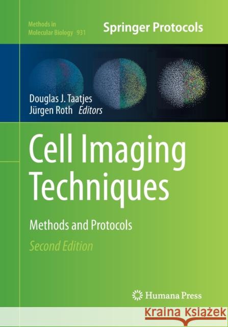
This contribution to the Methods in Molecular Biology collection has been updated to include new chapters covering colocalization and laser microdissection, among other emerging techniques. It features the series trademark of clear and reproducible protocols.
From the book reviews:
"The different techniques are described in deep details, the chapters having an abstract, introduction, materials, methods, and, last but not least, notes; each method is described step by step with very, very useful links to the notes. ... should be read not only by very specialized scientists, but also by those who usually work with classic microscopic methods, to open their minds and increase their appetite for more precise and complex measurements done on different level of complex biological structures." (Ioan I. Ardelean, Bulletin of Micro and Nanoelectrotechnologies, Vol. 5 (3-4), December, 2014)
"The goal is to provide seasoned microscopists with a list of protocols to add sophistication to their existing microscopy regimen. The intended audience is experienced researchers who are familiar with imaging in a core microscopy facility. ... this book is more useful to microscopists who routinely use microscopic techniques in their research and are knowledgeable about the recent advances in technology. ... an ideal reference for a core microscope facility. Each chapter is written independently by a different group of researchers and can stand alone." (Latha Malaiyandi, Doody's Book Reviews, December, 2013)
1. Digital Images are Data – and Should be Treated as Such
Douglas W. Cromey
2. Epi-fluorescence Microscopy
Donna J. Webb and Claire M. Brown
3. Live-Cell Migration and Adhesion Turnover Assays
Lacoste, J. Young, K. and Brown, C. M.
4. Multifluorescence Confocal Microscopy: Application for a Quantitative Analysis of HemostaticProteins in Human Venous Valves
Winifred E. Trotman, Douglas J. Taatjes, and Edwin G. Bovill
5. Colocalization Analysis in Fluorescence Microscopy
Jeremy Adler
6. A Time-Lapse Imaging Assay to Study Nuclear Envelope Breakdown
Sunita S. Shankaran, Douglas R. Mackay, and Katharine S. Ullman
7. Light Sheet Microscopy in Cell Biology
Raju Tomer, Khaled Khairy and Philipp J. Keller
8. Image-based High-throughput Screening for Inhibitors of Angiogenesis
Lasse Evensen, Wolfgang Link, and James B. Lorens
9. Intravital Microscopy to Image Membrane Trafficking in Live Rats
Andrius Masedunskas, Monika Sramkova, Laura Parente and Roberto Weigert
10. Imaging Non-fluorescent Nanoparticles in Living Cells with Wavelength-Dependent Differential Interference Contrast Microscopy and Planar Illumination Microscopy
Wei Sun, Lehui Xiao, and Ning Fang
11. Laser Scanning Cytometry. Principles and Applications: An Update
Piotr Pozarowski , Elena Holden, and Zbigniew Darzynkiewicz
12. Laser Capture Microdissection for Protein and NanoString RNA Analysis
Yelena Golubeva, Rosalba Salcedo, Claudius Mueller, Lance A. Liotta, and Virginia Espina
13. Viewing Dynamic Interactions of Proteins and a Model Lipid Membrane with Atomic Force Microscopy
Anthony S. Quinn, Jacob H. Rand, Xiao-Xuan Wu, and Douglas J. Taatjes
14. Mica Functionalization for Imaging of DNA and Protein-DNA Complexes with Atomic Force Microscopy
Luda S. Shlyakhtenko, Alexander A. Gall, and Yuri L. Lyubchenko
15. Measuring the Elastic Properties of Living Cells with Atomic Force Microscopy Indentation
Joanna L. MacKay and Sanjay Kumar
16. Atomic Force Microscopy Functional Imaging on Vascular Endothelial Cells
Lilia A. Chtcheglova and Peter Hinterdorfer
17. Porosome: The Secretory NanoMachine in Cells
Bhanu P. Jen18. Stereology and Morphometry of Lung Tissue
Christian Mühlfeld, Lars Knudsen, Matthias Ochs
19. A Novel Combined Imaging/Stereological Method for the Analysis of Human Sural Nerve Biopsies for Clinical Diagnosis
Michele A. VonTurkovich, Marilyn P. Wadsworth, William W. Pendlebury, and Douglas J. Taatjes
20. Correlative Light–Electron Microscopy as a Tool to Study In vivo Dynamics and uUtrastructure of Intracellular Structures
Elena V. Polishchuk, Roman S. Polishchuk and Alberto Luini
21. Photooxidation Technology for Correlative Light and Electron Microscopy
Claudia Meisslitzer-Ruppitsch, Clemens Röhrl, Carmen Ranftler, Herbert Stangl, Josef Neumüller, Margit Pavelka, Adolf Ellinger
22. Electron Microscopy of Endocytic Pathways
Carmen Ranftler, Peter Auinger, Claudia Meisslitzer-Ruppitsch, Adolf Ellinger, Josef Neumüller, Margit Pavelka
23. Morphological Analysis of Autophagy
Keisuke Tabata, Mitsuko Hayashi-Nishino, Takeshi Noda, Akitsugu Yamamoto, and Tamotsu Yoshimori
24. Cytochemical Detection of Peroxisomes and Mitochondria
Nina A. Bonekamp, Markus Islinger, Maria Gómez Lázaro, and Michael Schrader
25. Histochemical Detection of Lipid Droplets in Cultured Cells
Michitaka Suzuki, Yuki Shinohara, and Toyoshi Fujimoto
26. Environmental Scanning Electron Microscopy in Cell Biology
J. E. McGregor, L. T. L. Staniewicz, S. E. Guthrie (neé Kirk) and A. M. Donald 27. Environmental Scanning Electron Microscopy Gold Immunolabeling in Cell Biology F. Rosso, F. Papale and A. Barbarisi 28. High Pressure Freezing for Scanning Transmission Electron Tomography Analysis of Cellular Organelles Paul Walther, Eberhard Schmid, Katharina Höhn 29. MALDI Imaging Mass Spectrometry for Direct Tissue Analysis Stephan Meding and Axel Walch
27. Environmental Scanning Electron Microscopy Gold Immunolabeling in Cell Biology
F. Rosso, F. Papale and A. Barbarisi
28. High Pressure Freezing for Scanning Transmission Electron Tomography Analysis of Cellular Organelles
Paul Walther, Eberhard Schmid, Katharina Höhn
29. MALDI Imaging Mass Spectrometry for Direct Tissue Analysis
Stephan Meding and Axel Walch
Cell Imaging is rapidly evolving as new technologies and new imaging advances continue to be introduced. In the second edition of Cell Imaging Techniques: Methods and Protocols expands upon the previous editions with current techniques that includes confocal microscopy, transmission electron microscopy, atomic force microscopy, and laser microdissection. With new chapters covering colocalization analysis of fluorescent probes, correlative light and electron microscopy, environmental scanning electron microscopy, light sheet microscopy, intravital microscopy, high throughput microscopy, and stereological techniques. Written in the highly successful Methods in Molecular Biology™ series format, chapters include introductions to their respective topics, lists of the necessary materials and reagents, step-by-step, readily reproducible laboratory protocols, and tips on troubleshooting and avoiding known pitfalls
Authoritative and cutting-edge, Cell Imaging Techniques: Methods and Protocols, Second Edition is an easily accessible volume of protocols to be used with a variety of imaging-based equipment likely available in a core imaging facility.
1997-2026 DolnySlask.com Agencja Internetowa
KrainaKsiazek.PL - Księgarnia Internetowa


