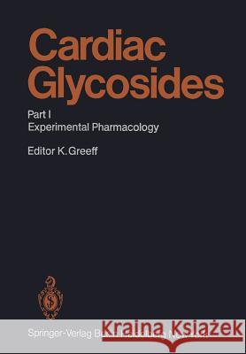
Cardiac Glycosides: Part I: Experimental Pharmacology » książka


Cardiac Glycosides: Part I: Experimental Pharmacology
ISBN-13: 9783642681653 / Angielski / Miękka / 2011 / 682 str.
Cardiac Glycosides: Part I: Experimental Pharmacology
ISBN-13: 9783642681653 / Angielski / Miękka / 2011 / 682 str.
(netto: 383,36 VAT: 5%)
Najniższa cena z 30 dni: 385,52
ok. 22 dni roboczych.
Darmowa dostawa!
Following the monographs by STRAUB (1924) and LENDLE (1935), this is the third contribution to the "Pharmacology of Cardiac Glycosides" within the Handbook of Experimental Pharmacology, which was founded by ARTHUR HEFFTER and con- tinued by WOLFGANG HEUBNER. Because of the need created by the length of time that had elapsed since LENDLE'S work, the editorial board requested the rapid ap- pearance of this 56th volume, which represents current knowledge of the pharma- cology and clinical pharmacology of cardiac glycosides. In order to avoid any delay, numerous authors were invited to contribute because shorter contributions take less time to prepare and are consequently more up-to-date. The disadvantage is that some overlap between certain chapters could not be avoided, despite the editor's efforts. Overlapping can, however, actually be useful, in that differing opinions may be provided and topical issues discussed from varying viewpoints. This re- minds the reader that scientific horizons in medicine should often be widened or revised. I would like to thank DR. ALANNA Fox and DR. K. ANANTHARAMAN for their help and advice in the revision of certain chapters. I am also grateful to Springer- Verlag, and particularly to MR. WINSTANLEY and MR. EMERSON, for their contribu- tion to the completion of this volume through translation and corrections. In con- clusion I would like to thank MRS. WALKER, MR. BISCHOFF, MRS. SEEKER, and MR. BERGSTEDT of Springer-Verlag for their helpful support.
1 Introduction and Remarks on the History of Cardiac Glycosides.- 2 Chemistry and Structure-Activity Relationships of Cardioactive Steroids.- A. Introduction.- B. Structure-Activity Relationships.- I. Uncertainties in Structure-Activity Relationships.- II. 3?-OH Group.- III. A–B Connection.- IV. C–D Connection.- V. Structure at C14 and C15.- VI. Side-Chain.- C. Influence of Additional Structural Modifications.- I. Halogens.- II. Branching at C3.- III. N-Analogs.- IV. Structure at C16.- V. Other Compounds.- D. Summary.- References.- Methods for the Determination of Cardiac Glycosides.- 3 Chemical and Chromatographic Methods.- A. Introduction.- B. Spectroscopic Procedures.- I Alkaline Reagents.- II. Acidic Reagents.- III. Fluorescence Spectroscopy.- IV. Quantitative Determination After Chromatography.- V. Quantitative Determination in Biologic Material.- C. Chromatographic Procedures.- I. Paper Chromatography and Thin Layer Chromatography.- II. Gas Chromatography.- III. Liquid Chromatography.- References.- 4 Use of Radioactively Labeled Glycosides.- A. Introduction.- B. Prerequisites for the Use of Isotope Techniques.- C. Production of Radioisotope Labeled Glycosides.- I. Biosynthesis.- II. Wilzbach Labeling.- III. Catalytic Exchange with Tritium Water.- IV. Reductive Tritiation.- V. Partial Synthetic Procedures.- D. Stability of the Radioactive Label.- E. Purity Testing of Labeled Glycosides.- F. Pharmacokinetic Investigations with Labeled Cardiac Glycosides.- G. Pharmacologic Investigations in Humans.- References.- 5 Radioimmunologic Methods.- A. Radioimmunoassay.- I. Basic Principles.- II. Antibodies.- 1. Immunogens and Immunization.- 2. Characterization.- III. Tracers.- 1. General Remarks.- 2. Conjugates for Labeling with 125I.- 3. Iodination.- IV. Standards.- V. Separation Methods.- VI. Assay Performances.- VII. Automation.- B. Enzyme Immunoassay.- I. Introduction.- II. Heterogeneous Enzyme Immunoassay.- III. Homogeneous Enzyme Immunoassay.- References.- 6 ATPase for the Determination of Cardiac Glycosides.- A. Introduction.- B. Preparation of ATPase.- C. Extraction Procedure from Biological Fluids.- D. Determination Based on Measurement of Enzyme Activity.- I. Measurement of the Hydrolysis of ATP.- II Inhibition of ATPase by Different Cardiac Glycosides.- III. Precision and Sensitivity of the Assay.- IV. Comparison of Results Obtained by ATPase Activity and Radioimmunoassay.- E. Determination Based on Isotope Displacement.- I. Binding Affinities of Different Cardiac Glycosides.- II. Precision and Sensitivity of the Assay.- III. Comparison of Results Obtained by Isotope Displacement and Other Assays.- F. Commentary.- I. Preparation of ATPase.- 1. ATPase Activity Assay.- 2. Isotope Displacement Assay.- II. Extraction Procedure.- 1. Dichlormethane.- 2. Chloroform.- III. Assay Procedure.- 1. ATPase Activity Assay.- 2. Isotope Displacement Assay.- References.- 7 Rubidium Uptake in Erythrocytes.- A. Introduction and Principle of the Method.- B. Factors Affecting the 86Rb Uptake of Human Erythrocytes.- I. Measurement of 86Rb-Activity.- II. Influences from Incubation Medium, Ion Concentrations, and pH.- 1. Influence of Rb+ Concentration and Specific Activity.- 2. Influence of Sodium Concentration.- 3. Influence of Calcium and Magnesium Concentration.- 4. Influence of pH During Incubation.- III. Influence of the Erythrocyte Preparation.- 1. Concentration of Erythrocytes in the Incubation Medium.- 2. Age of the Erythrocyte Preparation.- 3. Source of Erythrocyte Samples.- IV. Influence of Incubation Procedures on the 86REA.- 1. Incubation Temperature.- 2. Time of Incubation of Erythrocytes with 86Rb+.- 3. Influence of Preincubation of Erythrocytes with Digitalis.- V. Separation of Erythrocytes from Incubation Medium After Incubation.- C. Influence of Various Cardiac Glycosides, Genins, and Conjugates on the 86REA.- D. Specificity of the Inhibition of 86Rb Uptake.- I. Diverse Drugs.- II. Spironolactone.- III. Human Plasma.- E. Correlation of Activity of Cardiac Glycosides in 86REA and Cardioactivity.- F. Use of 86REA for Measurement in Body Fluids.- I. Plasma Glycoside Concentrations.- 1. Extraction Procedure.- 2. Preparation of the Extract.- 3. Preparation of Erythrocytes.- 4. Incubation Assay.- 5. Standard Curves.- 6. Calculation.- II. Glycoside Concentrations in Different Biological Media.- G. Comparison of Plasma Glycoside Measurements Using 86REA and Immunochemical Methods.- I. Determination of Plasma Digoxin.- II. Determination of Plasma Digitoxin.- H. Criticism of the Method as Used for Serum and Tissue Glycoside Concentration Determination.- I. Extraction.- II. Plasma Volume.- III. Biological Standard.- IV. Various Cardiac Glycosides.- V. Range of Discrimination.- VI. Use of the Method.- VII. Precision and Accuracy.- J. Plasma Concentrations of Cardiac Glycosides.- References.- Biological Methods for the Evaluation of Cardiac Glycosides.- 8 Evaluation of Cardiac Glycosides in the Intact Animal.- A. Introduction.- B. Toxicity as a Parameter of Biologic Efficacy.- I. Determination of the Lethal Dose in Anesthetized Animals by Intravenous Infusion Continued Until Cardiac Arrest.- 1. Cats.- 2. Guinea Pigs.- 3. Dogs.- 4. Pigs.- II. Determination of the Lethal Dose in Unanesthetized Animals.- 1. Frogs.- 2. Pigeons.- 3. Mice and Rats.- III. Factors Which Modify Toxicity.- 1. Anesthesia.- 2. Hypothermia.- 3. Hypoxia.- 4. Acidosis.- 5. Alkalosis.- 6. Age.- 7. Seasons.- 8. Autonomic Tone.- C. Sublethal Parameters of the Efficacy of Cardiac Glycosides.- I. The Inotropic Effect.- II. The Arrhythmogenic Effect.- III. The Kaliuretic Effect.- IV. Subacute Poisoning.- D. Determination of the Therapeutic Range.- I. Arrhythmogenic Dose and Lethal Dose.- II. Inotropic Dose and Lethal Dose.- III. Experimental Cardiac Failure.- E. Intestinal Absorption.- I. Comparison of Oral with Intravenous or Subcutaneous Efficacy in Unanesthetized Animals.- 1. By Determining the Lethal Dose.- 2. By Demonstrating ECG Changes.- 3. By Determining the Kaliuretic Effect in Rats.- 4. Tolerance Test with Acetylstrophanthidin After Oral Pretreatment.- II. Comparison of the Lethal Dose or Arrhythmogenic Dose by Intraduodenal and Intravenous Infusion in Anesthetized Animals.- III. Oral or Intraduodenal Pretreatment Followed by Determination of the Supplementary Dose in Anesthetized Animals.- IV. Determination of the Residue After Intraduodenal Administration.- V. Determination of Intestinal Absorption by Radiochemical or Radioimmunologic Methods.- VI. Determination of Hepatic Extraction.- F. Measurement of Cumulation and Duration of Action.- I. Repeated Administration of Sublethal Doses to Unanesthetized Experimental Animals.- II. Single Administration of a Sublethal Dose to Unanesthetized Animals Followed by Intravenous Titration Under Anesthesia.- III. Titration at Different Infusion Rates.- IV. Determination by Radiochemical or Radioimmunologic Methods.- References.- 9 The Use of the Isolated Papillary Muscle for the Evaluation of Positive Inotropic Effects of Cardioactive Steroids.- A. The Inotropic Potency.- B. Methodologic Considerations.- I. Selection and Preparation of Muscle.- II. Incubation Medium.- 1. Bicarbonate.- 2. Potassium.- 3. Calcium.- III. Temperature.- IV. Frequency of Contraction.- V. Stimulation Intensity.- VI. Length-Force Relationship and Plasticity.- VII. Stray Compliance.- VIII. The Two-Chambered Bath.- References.- 10 Evaluation of Cardiac Glycosides in Isolated Heart Preparations Other than Papillary Muscle.- A. Introduction.- B. Isolated Atrial Preparations.- I. Methods.- II. Evaluation of Effects.- 1. Qualitative Evaluation.- 2. Quantitative Evaluation.- 3. Interactions with Other Drugs.- C. Isolated Perfused Heart Preparation.- I. Method.- II. Quantitative Evaluation.- III. Interactions with Other Drugs.- D. Heart-Lung Preparations.- I. Method.- II. Therapeutic Effect and Therapeutic Range.- III. Interactions.- IV. Metabolic Characterization of Therapeutic Effects.- E. Isolated Frog Heart.- I. Method.- II. Application.- F. Embryonic Chicken Heart.- I. Method.- II. Application.- G. Isolated Cultures of Heart Cells.- I. Method.- II. Application.- References.- Mode of Action of Cardiac Glycosides.- 11 The Positive Inotropic Action of Cardiac Glycosides on Cardiac Ventricular Muscle.- A. Effects on the Time Course of the Isometric Contraction.- I. The Positive Klinotropic Effect.- II. Time to Peak Force and Relaxation Time.- III. Aftercontractions and Contracture.- B. Dependence on Extracellular Ion Concentrations.- I. Sodium Dependence.- II. The Influence of Potassium.- C. Frequency Dependence.- I. General Aspects of Drug Effects on the Frequency—Force Relationship of Cardiac Tissue.- 1. Ventricular Muscle.- 2. Atrial Muscle.- 3. The Atrium-Specific, Rested-State Contraction.- II. The Influence of Contraction Frequency on Steady-State Inotropic Effects.- 1. Ventricular Muscle.- 2. Atrial Muscle.- III. The Influence of Frequency on the Rate of Development of the Inotropic Effect.- 1. Ventricular Muscle.- 2. Atrial Muscle.- IV. On the Mechanism of the Frequency Dependence of Inotropic Steroid Action.- References.- 12 Influence of Cardiac Glycosides on Electrophysiologic Processes.- A. Introduction.- B. Electrophysiologic Background.- I. Ionic Current Components Underlying the Cardiac Action Potential.- 1. Working Myocardium.- 2. Specialized Conducting System.- II. Excitation-Contraction Coupling.- III. Electrical Coupling and Impulse Spread.- C. The Positive Inotropic Effect.- I. Modification of the Na+, K+-Pump.- 1. Correlation Between Pump Inhibition and Positive Inotropy.- 2. Correlation Between Pump Stimulation and Positive Inotropy.- II. Modification of the Slow Inward Current.- III. Calcium/Potassium Exchange Hypothesis.- D. Toxic Effects.- I. Circus Movement.- 1. Ionic Conductances.- 2. Passive Electrical Properties.- II. Alterations in Automaticity.- 1. Transient Depolarization.- 2. Ionic Basis of the Altered Automaticity.- 3. Current and Tension Fluctuations.- III. Influence on the Resting Tension.- E. Conclusions.- References.- 13 Influence of Cardiac Glycosides on Myocardial Energy Metabolism.- A. Introduction.- I. Myocardial Energy Metabolism.- 1. Energy Liberation.- 2. Energy Conservation.- 3. Energy Utilization.- II. General Biochemical Aspects of Myocardial Metabolism.- III. Cardiac Function as a Determinant of Myocardial Metabolism.- B. Influence of Cardiac Glycosides on Myocardial Energy Liberation.- I. Myocardial Oxygen Consumption.- II. Substrate Oxidation.- 1. Carbohydrate Metabolism.- 2. Fatty Acid Metabolism.- 3. Enzyme Activities.- C. Influence of Cardiac Glycosides on Myocardial Energy Conservation.- I. Myocardial Content of Energy-Rich Compounds.- II. Oxidative Phosphorylation.- D. Influence of Cardiac Glycosides on Myocardial Energy Utilization.- I. Utilization of Energy-Rich Compounds.- II. Myocardial Heat Production.- E. Additional Metabolic Effects.- F. Conclusions.- References.- 14 Effects of Cardiac Glycosides on Na+, K+-ATPase.- A. Introduction.- B. Binding of Cardiac Glycosides to Isolated Na+, K+-ATPase.- C. Kinetics and Stoichiometry of Glycoside Binding.- D. Effects of K+ on Glycoside Binding.- E. Glycoside Binding Sites and Release of Bound Glycosides.- F. Factors that Influence the Interaction of Cardiac Glycosides with Na+, K+-ATPase.- I. Chemical Structure of the Glycosides.- II. Source of the Enzyme.- III. Pathological Conditions, Temperature, and Membrane Lipids.- G. Consequences of Glycoside Binding in Cardiac Muscle.- I. Enzyme and Sodium Pump Activities.- II. Reserve Capacity of the Sodium Pump and Sodium Transients.- H. A Mechanism of Positive Inotropic Action.- J. Conclusions.- References.- 15 Influence of Cardiac Glycosides on their Receptor.- A. Introduction.- I. Definition of Receptors.- II. Binding Sites for Cardiac Glycosides.- Specific and Nonspecific Binding of Cardiac Glycosides.- B. Quantitative Aspects of Cardiac Glycoside-Receptor Interaction.- I. Quantitation of Ouabain-Binding Sites.- II. Correlation of Ouabain Binding and Na+, K+-ATPase Inhibition.- III. Quantitation of Ouabain-Binding Sites in Erythrocytes.- IV. Kinetics of Specific Ouabain-Receptor Binding.- V. Uniformity and Nonuniformity of Cardiac Glycoside Receptors.- Ouabain-Binding Sites in a Cell Membrane Preparation and in Contracting Ventricular Strips of Rat Heart.- VI. Correlation of Ouabain Binding and Increase in Force of Contraction.- VII. Dissociation Constants of the Ouabain-Receptor Complex and Sensitivity of Cardiac Glycosides.- VIII. Changes in Ouabain Receptor Density.- C. Qualitative Aspects of Cardiac Glycoside-Receptor Interaction.- I. Effects of Cations on Ouabain-Receptor Interaction.- II. Effect of Vanadate on Ouabain Binding.- III. Influence of pH and Temperature on Ouabain Binding.- D. Specificity of the Cardiac Glycoside Receptor.- I. Receptor Specificity for Cardiac Glycosides.- II. Other Substances that Bind to the Cardiac Glycoside Receptor.- III. Possible Application of Drug-Receptor Binding Studies in Experimental Pharmacology.- E. Conclusions.- References.- 16 Stimulation and Inhibition of the Na+, K+-Pump by Cardiac Glycosides.- A. Introduction.- B. Dose-Response Relationship.- C. Role of Duration of Treatment with Glycoside.- D. Molecular Requirements for Stimulation of the Pump.- E. Influence of Extracellular KC1 on Stimulation and Inhibition of the Pump by Ouabain.- F. Changes in Pump Activity and Inotropic Effect.- G. Receptor Sites Responsible for Stimulation and Inhibition of the Pump.- H. Concluding Remarks.- References.- 17 Influence of Cardiac Glycosides on Cell Membrane.- A. Function of Na+, K+-ATPase in Heart Muscle Cells.- B. Interaction of Cardiac Glycosides with Na+, K+-ATPase of Cardiac Tissue.- I. Na+-K+-ATPase as a Calcium Binding Partner.- II. Action of Cardiac Glycosides on Resting Cardiac Muscle.- C. Conclusions.- References.- 18 Influence of Cardiac Glycosides on Electrolyte Exchange and Content in Cardiac Muscle Cells.- A. Introduction.- B. Critical Evaluation of Factors Influencing the Validity of Myocardial Electrolyte Determinations.- C. Influence of Extracellular Electrolyte Composition on the Pharmacological Effect of Cardiac Glycosides.- I. Potassium.- II. Sodium.- III. Calcium.- D. Effect of Cardiac Glycosides on Transmembrane Ion Movements.- I. K+ and Na+ Fluxes in the Presence of Positive Inotropic Response to Cardiac Glycosides.- II. Ca2+ Fluxes in the Presence of Positive Inotropic Concentrations of Cardiac Glycosides.- III. K+, Na+, and Ca2+ Exchange in the Presence of Toxic Concentrations of Cardiac Glycosides.- E. Effect of Cardiac Glycosides on Intracellular Electrolyte Content.- I. Influence of Cardiac Glycosides on Myocardial K+ and Na+.- II. Influence of Cardiac Glycosides on Myocardial Calcium Content.- F. Effect of Cardiac Glycosides on Subcellular Calcium Storage Sites.- I. Sarcoplasmic Reticulum.- H. Mitochondria.- G. Conclusions.- References.- 19 Effects of Cardiac Glycosides on Myofibrils.- A. Introduction.- B. Molecular Aspects of Myofibrillar Proteins.- I. Contractile Mechanism.- II. Regulation of Contraction.- C. Effect of Cardiac Glycosides on Myofibrillar Function.- I. Calcium.- II. Phosphorylation.- D. Conclusions and Outlook.- References.- 20 Substances Possessing Inotropic Properties Similar to Cardiac Glycosides.- A. Introduction.- B. Na+, K+-ATPase Inhibitors.- I. Cassaine.- II. Prednisolone-bis-Guanylhydrazone (PBGH).- III. Benzylaminodihydrodimethoxyimidazoisoquinoline Hydrochloride (BIIA).- IV. Sulfhydryl Blocking Agents.- V. Monovalent Cations.- VI. Vanadate.- VII. Other Na+, K+-ATPase Inhibitors.- C. Substances that Enhance Sodium Influx.- D. Conclusions.- References.- Non-Cardiac Effects of Cardiac Glycosides.- 21 Effects of Cardiac Glycosides on Central Nervous System.- A. Introduction.- B. Central Vomiting.- C. Respiration.- D. Central Parasympathetic Activity.- E. Central Sympathetic Activity.- F. Central Excitation.- G. Visual Symptoms.- H. Neurotoxicity in Humans.- References.- 22 Effects of Cardiac Glycosides on Vascular System.- A. Introduction.- B. Systemic Arterioles in Experimental Animals.- I. Total Vascular Resistance.- II. Regional Vascular Resistance.- C. Systemic Veins in Experimental Animals.- I. Total Systemic Venous Tone.- II. Hepatic Venous Tone.- D. Systemic Arterioles in Patients.- E. Systemic Arterioles and Veins in Normal Human Subjects.- I. Total and Regional Vascular Effects.- II. Direct Vascular Actions.- F. Systemic Arterioles and Veins in Patients with Congestive Heart Failure.- I. Total and Regional Vascular Effects.- II. Mechanisms of Vascular Action.- G. Significance of Digitalis-Induced Vasomotor Changes.- I. Normal Versus Heart Failure.- II. Influence on Cardiac Output.- III. Influence on Cardiac Preload and Energetics.- IV. Coronary Heart Disease.- V. Acute Pulmonary Edema.- VI. Compensated Heart Failure.- H. Conclusions.- References.- 23 Effects of Cardiac Glycosides on Skeletal Muscle.- A. Introduction.- B. Basic Differences Between Skeletal and Heart, and Red and White Muscle.- I. Morphological Structure.- II. Electrophysiologic Properties.- III. The Membrane Enzyme Na+, K+-ATPase.- IV. Binding and Accumulation.- V. Red and White Muscle.- C. Influences on Force of Contraction of Skeletal Muscle.- I. In Situ Experiments.- II. In Vitro Experiments.- D. Influences on Skeletal Muscle Electrolyte Content.- E. Influence on the Electrophysiologic Parameters of Skeletal Muscle.- References.- 24 Effects of Cardiac Glycosides on Autonomic Nervous System and Endocrine Glands.- A. Autonomic Effects of Cardiac Glycosides.- I. Effects on the Sympathetic Nervous System.- II. Effects on the Parasympathetic Nervous System.- III. Effects on the Gastrointestinal System.- IV. Respiratory Effects.- B. Endocrine Effects of Cardiac Glycosides.- I. Estrogenic Effects and Gynecomastia.- II. Adrenocortical Hormones.- III. Thyroid Hormones.- IV. Other Endocrine Glands.- References.- 25 Effects of Cardiac Glycosides on Kidneys.- A. Introduction.- B. Effects on Renal Hemodynamics.- C. Effects on Renal Excretion of Solutes and Water.- I. Excretion of Sodium.- II. Excretion of Potassium.- III. Excretion of Other Electrolytes and Nonelectrolytes.- 1. Calcium and Magnesium Reabsorption.- 2. Hydrogen Ion Excretion.- 3. Chloride and Phosphate Reabsorption.- 4. Glucose Reabsorption.- 5. Excretion of Organic Anions.- 6. Excretion of Organic Cations.- IV. Excretion of Water.- V. Quantitative Differences in the Diuretic Activity of Cardiac Glycosides.- D. Site of Renal Action.- E. Mechanism of Tubular Action of Cardiac Glycosides.- I. Distribution and Localization of Na+, K+-ATPase in the Kidney.- II. Binding of Cardiac Glycosides to Renal Tissue.- III. Correlation Between Inhibition of Na+, K+-ATPase and Tubular Reabsorption of Solutes and Fluid.- IV. Transepithelial Electric Potential Difference.- V. Effects on Oxygen Consumption and Renal Metabolism.- References.- Author Index.
1997-2026 DolnySlask.com Agencja Internetowa
KrainaKsiazek.PL - Księgarnia Internetowa


