Termin realizacji zamówienia: ok. 22 dni roboczych.
Darmowa dostawa!
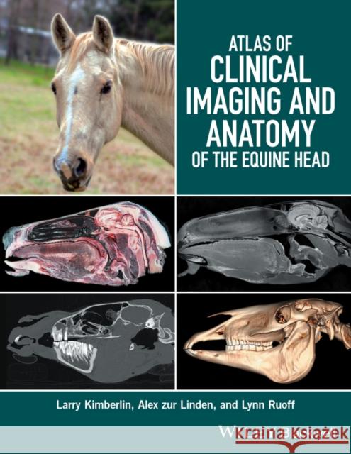
Atlas of Clinical Imaging and Anatomy of the Equine Head presents a clear and complete view of the complex anatomy of the equine head using cross-sectional imaging.
Provides a comprehensive comparative atlas to structures of the equine head
Pairs gross anatomy with radiographs, CT, and MRI images
Presents an image-based reference for understanding anatomy and pathology
Covers radiography, computed tomography, and magnetic resonance imaging
Bibliografia Glosariusz/słownik Wydanie ilustrowane
"Atlas of Clinical Imaging and Anatomy of the Equine Head is a comprehensive reference of the cross–sectional anatomy of the head of equids that features photographs of gross sections, CT images, and MRI scans of the head in transverse, sagittal, and dorsal planes. The photographs of gross–section preparations are excellent, and most anatomic features are readily identifiable. Furthermore, the anatomic labels are exhaustive"...... "The provided images should enable readers to recognize most anatomic structures on other MRI pulse sequences"......"In addition to the anatomic atlas, the book contains a well–written introduction that briefly explains the basic principles of CT and MRI interpretation and considerations for image acquisition. Readers unfamiliar with CT and MRI will gain a superficial understanding of what influences tissue appearance in images acquired by the use of those modalities, and appropriate references are provided for readers who wish to learn more. This book will be useful for radiologists, surgeons, internists, and other practitioners who occasionally need to acquire diagnostic images of the heads of horses"
(Reviewed by Derek Cissell, VMD, PhD, DACVR, University of California–Davis, Davis, Calif 15th June 2017 AVMA)
Introduction: General Presentation of Atlas, vi
1 Overview of CT and MRI of the Equine Head, 1
2 Clinical and Surgical Anatomy of the Equine Head: Transverse Sections, 9
3 Clinical and Surgical Anatomy of the Equine Head: Sagittal Sections, 85
Brain sagittal close–up, 100
4 Clinical and Surgical Anatomy of the Equine Head: Dorsal Sections, 115
Glossary, 143
References, 145
Index, 147
Larry Kimberlin, DVM, FAVD, CVPP, is the owner of Northeast Texas Veterinary Dental Center in Greenville, Texas, USA.
Alex zur Linden, DVM, DACVR, is Assistant Professor of Radiology at Ontario Veterinary College at the University of Guelph in Ontario, Canada.
Lynn Ruoff, DVM is Clinical Associate Professor at Texas A&M University s College of Veterinary Medicine in College Station, Texas, USA.
Atlas of Clinical Imaging and Anatomy of the Equine Head presents a clear and complete view of the complex anatomy of the equine head using cross–sectional imaging. The gross anatomy of a one–centimeter section of the equine head is compared to identical slices in CT and MRI in the transverse, sagittal, and dorsal planes. To aid in the identification of clinically important structures, the book covers oral, dental, nasal, sinus, ophthalmic, auricular, laryngeal, hyoid apparatus and tongue structures.
The atlas offers more than 300 gross photographs, radiographs, CT images, and MRI images, with all structures indicated using color–coded labels. Veterinary students, equine practitioners, surgeons and imaging specialists who wish to foster a clear understanding of the anatomy of the structures involved in the equine head will find Atlas of Clinical Imaging and Anatomy of the Equine Head an essential resource.
Key features
Provides a comprehensive comparative atlas to structures of the equine head
Pairs gross anatomy with radiographs, CT, and MRI images
Presents an image–based reference for understanding anatomy and pathology
Covers radiography, computed tomography, and magnetic resonance imaging