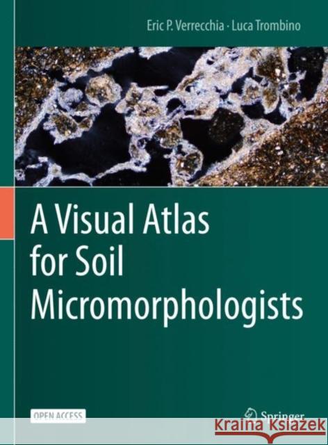
A Visual Atlas for Soil Micromorphologists » książka


A Visual Atlas for Soil Micromorphologists
ISBN-13: 9783030678050 / Angielski / Twarda / 2021 / 192 str.
A Visual Atlas for Soil Micromorphologists
ISBN-13: 9783030678050 / Angielski / Twarda / 2021 / 192 str.
(netto: 192,11 VAT: 5%)
Najniższa cena z 30 dni: 192,74
ok. 16-18 dni roboczych.
Darmowa dostawa!
Chapter 1. The multiscalar nature of soils.- Chapter 2. History of micromorphology.- Chapter 3. Observation and sampling of soils.- Chapter 4. How to make thin sections.- Chapter 5.The polarised light microscope.- Chapter 6. Other techniques of observation.- Chapter 7. Electron and energy imaging.- Chapter 8. Colours of minerals.- Chapter 9. The micromorphological approach.- Chapter 10. Concept of fabric.- Chapter 11. Multiscalar approach to fabric.- Chapter 12. Basic distribution patterns.- Chapter 13. c/f related distributions I.- Chapter 14. c/f related distributions II.- Chapter 15. Aggregates and aggregation.- Chapter 16. Degree of separation and accommodation of aggregates.- Chapter 17. The nature of voids.- Chapter 18. Morphology of voids I.- Chapter 19. The morphology of voids II.- Chapter 20. Microstructure I.- Chapter 21. Microstructure II.- Chapter 22. Mineral and organic constituents.- Chapter 23. Particle size and sorting.- Chapter 24. Shape of grains: equidimensionality.- Chapter 25. Shape of grains: roundness and sphericity.- Chapter 26. Basalt, granite, and gabbro.- Chapter 27. Schist, gneiss, and amphibolite.- Chapter 28. Quartzite and marble.- Chapter 29. Calcium-bearing sedimentary rocks.- Chapter 30. Sand and sandstone.- Chapter 31. Mineral grains in the soil I: quartz and chalcedony.- Chapter 32. Mineral grains in the soil II: feldspar and mica.- Chapter 33. Mineral grains in the soil III: inosilicates and nesosilicates.- Chapter 34. Mineral grains in the soil IV: carbonates.- Chapter 35. Mineral grains in the soil V: chlorides and sulfates.- Chapter 36. Biominerals I.- Chapter 37. Biominerals II.- Chapter 38. Biominerals III.- Chapter 39. Anthropogenic features I.- Chapter 40. Anthropogenic features II.- Chapter 41. Organic matter I.- Chapter 42. Organic matter II.- Chapter 43. Humus.- Chapter 44. Micromass.- Chapter 45. B-fabric I.- Chapter 46. B-fabric II.- Chapter 47. Imprints of pedogenesis.- Chapter 48. Iron- and manganese-bearing nodules.- Chapter 49. Carbonate nodules.- Chapter 50. Polygenetic nodules.- Chapter 51. Nodules: morphology and border shape.- Chapter 52. Nodules: orthic, anorthic, disorthic.- Chapter 53. Crystals and crystal intergrowths.- Chapter 54. Impregnations.- Chapter 55. Depletions.- Chapter 56. Coatings with clays I.- Chapter 57. Coatings with clays II.- Chapter 58. Micropans, coarse coatings, cappings, and crusts.- Chapter 59. Hypocoatings and quasicoatings: amorphous.- Chapter 60. Coatings and hypocoatings: crystalline.- Chapter 61. Mineral infillings.- Chapter 62. Mineral infillings of biological origin.- Chapter 63. Pedoturbations.- Chapter 64. Faecal pellets.- Chapter 65. Dung and vertebrate excrements.- Chapter 66. Composite pedogenic features.- Chapter 67. Uncommon features.- Chapter 68. Pedofeatures and soil processes.- Chapter 69. Clay dynamics I - Translocation.- Chapter 70. Clay dynamics II - Swelling.- Chapter 71. Water dynamics..- Chapter 72. Carbonate and gypsum dynamics.- Chapter 73. Processes involving iron oxyhydroxides.- Chapter 74. Biogeochemical processes I.- Chapter 75. Biogeochemical processes II.- Chapter 76. The future of soil micromorphology.- Chapter 77. Beyond the two dimensions.- Chapter 78. The prospect of chemical imaging
Dr. Eric P. Verrecchia is a full professor of Biogeosciences at the Institute of Earth Surface Processes, University of Lausanne (Switzerland). He specializes in geopedology and biogeochemistry of the terrestrial carbon and calcium cycles. Awarded with a Marie-Curie-Fellowship in 1994-95, he joined Prof. G. Stoops laboratory of soil micromorphology in Gent (Belgium), where he was introduced to the microscopic approach of soils. Since then, he has been teaching and applying this technique, by coupling it with biogeochemical methods, to soils from the tropics and the temperate zone, particularly in carbonate-rich environments.
This open access atlas is an up-to-date visual resource on the features and structures observed in soil thin sections, i.e. soil micromorphology. The book addresses the growing interest in soil micromorphology in the fields of soil science, earth science, archaeology and forensic science, and serves as a reference tool for researchers and students for fast learning and intuitive feature and structure recognition. The book is divided into six parts and contains hundreds of images and photomicrographs. Part one is devoted to the way to sample properly soils, the method of preparation of thin sections, the main tool of soil micromorphology (the microscope), and the approach of soil micromorphology as a scientific method. Part two focuses on the organisation of soil fragments and presents the concept of fabric. Part three addresses the basic components, e.g. rocks, minerals, organic compounds and anthropogenic features. Part four lists all the various types of pedogenic features observed in a soil, i.e. the imprint of pedogenesis. Part five gives interpretations of features associated with the main processes at work in soils and paleosols. Part six presents a view of what the future of soil micromorphology could be. Finally, the last part consists of the index and annexes, including the list of mineral formulas. This atlas will be of interest to researchers, academics, and students, who will find it a convenient tool for the self-teaching of soil micromorphology by using comparative photographs.
1997-2026 DolnySlask.com Agencja Internetowa
KrainaKsiazek.PL - Księgarnia Internetowa


