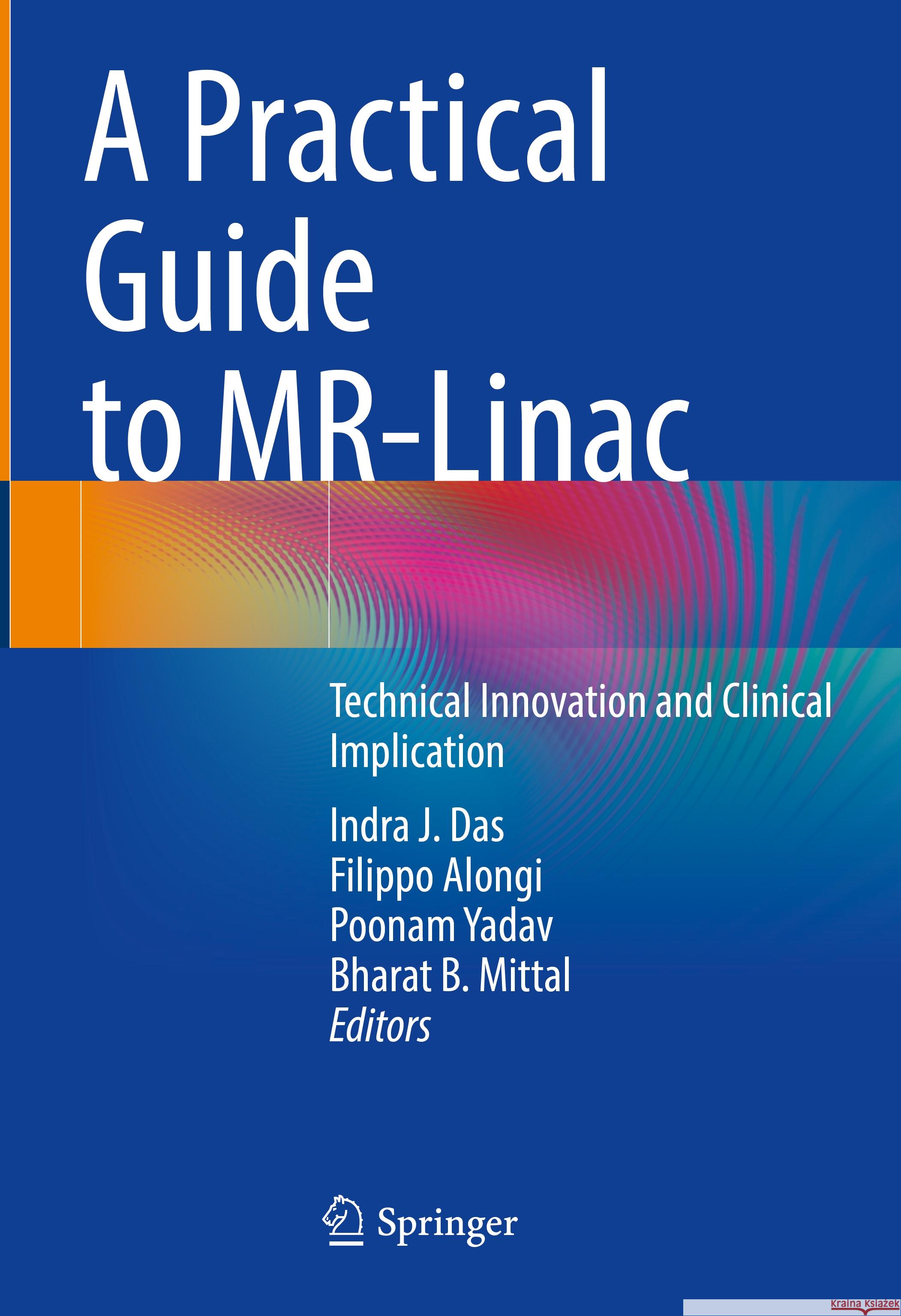
A Practical Guide to Mr-Linac: Technical Innovation and Clinical Implication » książka


A Practical Guide to Mr-Linac: Technical Innovation and Clinical Implication
ISBN-13: 9783031481642 / Angielski
A Practical Guide to Mr-Linac: Technical Innovation and Clinical Implication
ISBN-13: 9783031481642 / Angielski
(netto: 613,40 VAT: 5%)
Najniższa cena z 30 dni: 616,85
ok. 22 dni roboczych.
Darmowa dostawa!
I.General 1. Introduction: 2. Role of MRI in Radiation Oncology: a. Multimodality imaging
b. Image Fusion
c. Contouring issues
d. Pre and post treatment imaging
3. Clinical necessity & Patient Selection
e. Selectionf. IRB and regulatory issues
g. Coaching and expectation
h. Patient satisfaction
4. MRL: Education and Training a. Physicians: Radiation Oncologistb. Physicists
c. Dosimetrists
d. Radiation Therapists and Nurses
e. Patient
II. MRL Physics and Technology 5. Imaging sequence (min 2 authors from Unity and View ray) 6. Motion Management and Tracking (Unity and View ray) 7. Synthetic CT and dose calculation 8. Treatment planning (authors from View ray & Unity each) a. TPS and algorithm consideration b. Contouring c. DVH constraints d. Optimization e. Dose calculation f. Evaluation, accuracy Systems Each MRL technology chapters should have nearly identical layout as described below with subheading; i. Planning, Request for Proposal (RFP) ii. Selection criterion iii. Architectural & Structural Planning iv. Cost and economic analysis for rural vs urban center v. Construction and installation time line vi. Acquisition of MR-compatible equipment and devices vii. Acceptance and commissioning viii. Setting daily, PSQA, monthly and annual QA ix. QA, Calibration and Imaging x. Safety consideration xi. IT and other consideration 9. View Ray system 10. Elekta Unity System 11. Aurora, Magneton system III. Clinical Each clinical site chapters should have nearly identical lay out as described below with subheading; i. Disease overview ii. Overcoming Challenges with Traditional Approaches iii. Contouring, PTV and OAR iv. DVH constraints v. Dose escalation vi. On table adaptation vii. Evaluation viii. Outcome ix. Other 12. Oligometastatic 13. Pancreas 14. Liver 15. Lung 16. Prostate 17. Gynecology 18. Breast 19. Spine 20. Brain 21. Head and neck 22. Sarcoma IV. Future and outlook 23. New MRL System a. Sydney project 24. Low field Imaging a. Imaging parameters used b. Integration in clinical unit c. Clinical consequences 25. AI, Radiomics and Texture analysis 26. MRL and biomarkersIndra J Das, PhD is Professor of Radiation Oncology at Northwestern University Feinberg School of Medicine, Chicago, IL, USA. He was recently selected by the AAPM to be the 2022 recipient of the Edith H. Quimby Award for Lifetime Achievement in Medical Physics. He is co-editor of Basic Radiotherapy Physics and Biology, 2e (2021).
Poonam Yadav, PhD is Associate Professor of Radiation Oncology at Northwestern University Feinberg School of Medicine, Chicago, IL, USA.
Bharat B. Mittal, MD is Chair and the William N. Brand, MD, Professor of the Department of Radiation Oncology, Northwestern University Feinberg School of Medicine, Chicago, IL, USA.
This book offers a detailed guide to MR-Linac, a unique and fast growing radiation treatment modality. MR-linac is new technology that is a fusion of an MRI and a linear accelerator on the same gantry. It can change both target volume delineation and tumor visualization in real time using MR-cine images and treatment. Tumor location changes moment to moment as radiation is delivered, but this cannot be visualized in current radiation therapy practices. This new and rapidly growing technology can provide adaptive therapy that was not possible before.
This book presents current knowledge on MR-linac technology, clinical practices, and ultimately patient outcome where dose escalation is not possible due to limiting normal tissue structures in the vicinity of tumor. There are two commercial MR-linac machines under consideration and both will be covered in detail. The book is divided into four sections. The first gives a general introduction to MR-Linac, covering the role of MRI in radiation oncology, the clinical necessity of this technology, and patient selection. The next section details the physics and technology of MR-Linac, covering image sequence, motion management, and treatment planning. Section three offers the clinical applications of MR-Linac and is divided by body area, including lung, prostate, and breast. Finally, the fourth section looks to the future and what this technology can mean for radiation oncology.This is an ideal guide for radiation oncologists, medical physicists, and relevant trainees.1997-2026 DolnySlask.com Agencja Internetowa
KrainaKsiazek.PL - Księgarnia Internetowa


