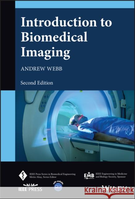
Introduction to Biomedical Imaging » książka


Introduction to Biomedical Imaging
ISBN-13: 9781119867715 / Angielski / Twarda / 2022 / 300 str.
Introduction to Biomedical Imaging
ISBN-13: 9781119867715 / Angielski / Twarda / 2022 / 300 str.
(netto: 554,94 VAT: 5%)
Najniższa cena z 30 dni: 580,29
ok. 30 dni roboczych.
Darmowa dostawa!
Preface xvIntroduction xixAbout the Companion Website xxxi1 Image and Imaging System Characteristics 11.1 General Image and Imaging System Characteristics 11.2 Concept of Spatial Frequency 21.3 Spatial Resolution 31.3.1 Imaging System Point Spread Function 41.3.2 Imaging System Resolving Power 51.3.3 Imaging System Modulation Transfer Function 61.4 Signal-to-Noise Ratio 71.5 Contrast-to-Noise Ratio 91.6 Signal Digitization: Dynamic Range and Resolution 91.7 Post-acquisition Image Filtering 111.8 Assessing the Clinical Impact of Improvements in System Performance 121.8.1 The Receiver Operating Characteristic Curve 131.A.1 Fourier Transforms 141.A.2 Fourier Transforms of Time Domain and Spatial Frequency Domain Signals 151.A.3 Useful Properties of the Fourier Transform 16Exercises 17References 19Further Reading 202 X-ray Imaging and Computed Tomography 232.1 General Principles of Imaging with X-rays 232.2 X-ray Production 252.2.1 The X-ray Tube 252.2.2 The X-ray Energy Spectrum 292.3 Interactions of X-rays with Tissue 322.3.1 Compton Scattering 332.3.2 The Photoelectric Effect 342.4 Linear and Mass Attenuation Coefficients of X-rays in Tissue 362.5 Instrumentation for Planar X-ray Imaging 382.5.1 Collimator 382.5.2 Anti-scatter Grid 382.6 Digital X-ray Detectors 402.7 X-ray Image Characteristics 422.7.1 Signal-to-Noise 422.7.2 Spatial Resolution 442.7.3 Contrast-to-Noise 452.8 X-ray Contrast Agents 462.8.1 Contrast Agents for the Gastrointestinal Tract 462.8.2 Iodine-Based Contrast Agents 462.9 X-ray Imaging Methods 472.9.1 X-ray Fluoroscopy 482.9.2 Digital Subtraction Angiography 482.10 Clinical Applications of X-ray Imaging 492.10.1 Digital Mammography 492.10.2 Abdominal X-ray Scans 502.11 Computed Tomography 512.12 CT Scanner Instrumentation 532.12.1 Beam Filtration 552.12.2 Detectors for Computed Tomography 562.13 Image Processing for Computed Tomography 572.13.1 Filtered Backprojection (FBP) Techniques 572.13.2 Fan-Beam and Spiral Reconstructions 612.14 Iterative Algorithms 632.15 Radiation Dose 652.16 Spectral/Dual Energy CT 662.17 Photon-Counting CT 692.18 Cone Beam, Mobile, and Portable CT Units 712.19 Clinical Applications of Computed Tomography 722.19.1 Head and Neurovascular Scans 722.19.2 Pulmonary Disease 732.19.3 Abdominal Imaging 732.19.4 Cardiovascular Imaging 74Exercises 75References 81Further Reading 823 Nuclear Medicine 853.1 General Principles of Nuclear Medicine 853.2 Radioactivity and Radiotracer Half-life 873.3 Common Radiotracers Used for SPECT 893.4 The Technetium Generator 903.5 The Distribution of Technetium-Based Radiotracers within the Body 923.6 Instrumentation for SPECT and SPECT/CT 943.6.1 Collimators 943.6.2 Scintillation Crystal and Photomultiplier Tube-Based Detectors 983.6.3 The Anger Position Network and Pulse Height Analyzer 1003.6.4 Solid-State Detectors and Specialized Cardiac Scanners 1023.7 Image Reconstruction 1033.7.1 Attenuation Correction 1043.7.2 Scatter Correction 1053.8 Image Characteristics 1063.8.1 Signal-to-Noise 1063.8.2 Spatial Resolution 1073.8.3 Contrast-to-Noise 1073.9 Clinical Applications of SPECT 1073.9.1 Brain Imaging 1083.9.2 Bone Scanning and Tumor Detection 1083.9.3 Cardiac Imaging 1103.9.4 The Respiratory System 1103.9.5 The Liver and Reticuloendothelial System 1123.10 Positron Emission Tomography 1133.11 Radiotracers Used for PET 1153.12 Instrumentation for PET 1163.12.1 Scintillation Crystals and Detector Electronics 1173.13 Image Reconstruction 1183.13.1 Annihilation Coincidence Detection and Removal of Accidental Coincidences 1193.13.2 Attenuation Correction 1203.13.3 Scatter Correction 1203.13.4 Dead-Time Correction 1203.14 Image Characteristics 1213.14.1 Spatial Resolution 1213.14.2 Signal-to-Noise 1213.14.3 Contrast-to-Noise 1223.15 Acquisition Methods for PET 1223.16 Total Body PET Systems 1223.17 Clinical Applications of PET/CT 1243.17.1 Body Oncology 1243.17.2 Brain Imaging 1253.17.3 Cardiac Imaging 125Exercises 126References 131Further Reading 1324 Ultrasound Imaging 1354.1 General Principles of Ultrasound Imaging 1354.2 Wave Propagation and Acoustic Impedance 1374.3 Wave Reflection 1394.4 Energy Loss Mechanisms in Tissue 1424.4.1 Scattering 1424.4.2 Absorption 1434.4.3 OverallWave Attenuation 1454.5 Instrumentation 1454.5.1 Transducer Construction 1464.5.2 Transducer Arrays 1494.5.2.1 Linear Sequential Array 1514.5.2.2 Curvilinear/Convex Sequential Array 1514.5.2.3 Linear-Phased Array 1524.6 Signal Detection and Processing 1534.6.1 Time Gain Compensation 1534.6.2 Receive Beam Forming 1544.7 Diagnostic Scanning Modes 1554.7.1 A-Mode, M-Mode, and B-Mode Scans 1554.7.2 Three-Dimensional Imaging 1564.7.3 Compound Imaging 1564.7.4 Other Transmit and Receive Beamforming Techniques 1584.8 Image Characteristics 1584.8.1 Signal-to-Noise 1584.8.2 Spatial Resolution 1594.8.2.1 Axial Resolution 1594.8.2.2 Lateral Resolution 1604.8.3 Contrast-to-Noise 1614.9 Artifacts in Ultrasound Imaging 1614.10 Blood Velocity Measurements Using Ultrasound 1634.10.1 The Doppler Effect 1634.10.2 Pulsed-Mode Doppler Measurements 1644.10.3 Color Doppler/B-mode Duplex and Triplex Imaging 1674.10.4 ContinuousWave Doppler (CWD) Measurements 1684.11 Ultrasound Contrast Agents 1694.11.1 Harmonic and Pulse Inversion Techniques 1714.11.2 Super-Resolution in Ultrasound Imaging 1724.12 Safety and Bioeffects in Ultrasound Imaging 1744.13 Point-of-Care Ultrasound Systems 1754.14 Clinical Applications of Ultrasound 1764.14.1 Obstetrics and Gynecology 1764.14.2 Breast Imaging 1764.14.3 Musculoskeletal Structure 1774.14.4 Abdominal 178Exercises 179References 185Further Reading 1865 Magnetic Resonance Imaging 1895.1 General Principles of MRI Acquisition and Hardware 1895.2 Nuclear Magnetization 1915.2.1 Quantum Mechanical Description 1915.2.2 Classical Description 1955.2.3 Hydrogen Nuclei inWater and Lipid 1975.2.4 Radiofrequency Pulses and the Creation of Transverse Magnetization 1975.2.5 Signal Detection and Fourier Transformation 1995.3 T1 and T2 Relaxation Mechanisms and Tissue Relaxation Times 2005.3.1 Tissue-Dependent Relaxation Times 2025.3.2 Measurement of T1 and T2: Inversion-Recovery and Spin-Echo Sequences 2045.4 The MR Free Induction Decay 2065.5 Magnetic Resonance Imaging 2075.5.1 Spatial Localization 2075.5.2 Imaging Concepts 2095.5.2.1 Slice Selection 2105.5.2.2 Phase-encoding 2125.5.2.3 Frequency-encoding 2145.5.2.4 The k-Space Formalism and Image Reconstruction 2145.6 Imaging Sequences and Techniques 2175.6.1 Multislice Gradient-Echo Sequences 2175.6.2 Multislice Spin-Echo and Turbo-Spin-Echo Sequences 2195.6.3 Three-Dimensional Gradient-Echo and Spin-Echo Sequences 2215.6.4 Proton Density, T1-, T2-, and T*2 -Weighted Sequences 2225.6.5 Lipid Suppression Techniques 2235.7 MRI Contrast Agents 2265.8 Advanced Sequences 2285.8.1 Magnetic Resonance Angiography 2285.8.2 Diffusion-Weighted Imaging with Echo Planar Readout 2305.8.3 In Vivo Localized Spectroscopy 2325.8.4 Functional MRI 2335.9 Instrumentation 2355.9.1 Magnet Design 2355.9.1.1 Clinical Superconducting Magnets 2355.9.1.2 Very High Field Magnets 2385.9.1.3 High-Temperature Superconductors 2395.9.1.4 Mid- and Low-Field Magnets 2395.9.2 Magnetic Field Gradient Coils 2405.9.3 Radiofrequency Coils 2445.9.3.1 Transmit Coil 2445.9.4 Receiver Coil Array 2445.9.5 Receiver Electronics 2465.10 Image Reconstruction from Undersampled Data 2475.10.1 Parallel Imaging Using an Array of Receiver Coils 2485.10.2 Compressed Sensing 2505.11 Image Characteristics 2525.11.1 Signal-to-Noise 2525.11.2 Spatial Resolution 2545.11.3 Contrast-to-Noise 2545.12 Image Artifacts 2555.13 RF Safety Considerations 2565.14 Clinical Applications of MRI 2575.14.1 Neurological 2585.14.2 Body Imaging 2595.14.3 Musculoskeletal 2595.14.4 Cardiac 259Exercises 262References 274Further Reading 2776 Optical Imaging 2796.1 General Properties of Optical Imaging Methods 2796.2 Propagation of Light Through Tissue 2816.3 Body Emissivity Techniques - Infrared Thermography 2846.4 Direct Imaging with Visible Light 2856.4.1 Fundus Photography 2856.4.2 Scheimpflug Camera 2876.5 Optical Coherence Tomography (OCT) 2886.5.1 Basic Principles of Interferometry 2896.5.2 Instrumentation for OCT 2916.5.2.1 Light Sources 2916.5.2.2 Beam-Splitter 2926.5.2.3 Photodetectors 2926.5.3 Image Characteristics of OCT 2926.5.4 OCT Angiography 2946.5.5 Clinical Applications of OCT 2956.6 Fluorescence-Guided Surgery (FGS) 2966.6.1 Principle of Fluorescence 2966.6.2 Fluorescent Probes 2966.6.3 Instrumentation for Fluorescence Imaging 2976.6.4 Clinical Applications of Fluorescence-Guided Surgery 2986.7 Near-Infrared Spectroscopy (NIRS) and Diffuse Optical Tomography (DOT) 2996.7.1 Principle of NIRS 2996.7.2 Instrumentation for NIRS 3016.7.3 Principle of DOT 3016.7.4 Clinical Applications of DOT 3026.8 Photoacoustic Imaging (PAI) 3036.8.1 Principles of PAI 3036.8.2 Photoacoustic Microscopy and Photoacoustic Computed Tomography 3046.8.3 Instrumentation for PAI 3056.8.4 Clinical Applications of PAI 305References 306Further Reading 3087 Artificial Intelligence 3117.1 Artificial Intelligence in Biomedical Imaging 3117.2 Artificial Intelligence, Machine Learning, Deep Learning, and Neural Networks 3127.2.1 Neural Networks 3137.3 Deep Learning in Image Reconstruction 3177.4 Convolutional Neural Networks (CNNs) 3187.5 Artificial Intelligence in X-ray and CT 3217.5.1 Image Reconstruction 3217.5.2 Clinical Applications 3227.6 Artificial Intelligence in SPECT and PET 3237.6.1 Image Reconstruction 3237.6.2 Clinical Applications 3247.7 Artificial Intelligence in Ultrasound 3247.7.1 Improved Data Acquisition 3257.7.2 Image Post-processing 3267.7.3 Image Analysis and Clinical Applications 3267.8 Artificial Intelligence in MRI 3267.8.1 Image Reconstruction 3267.8.2 Clinical Applications 3287.9 Artificial Intelligence in Optical Imaging 3297.10 AI and Radiomics 3297.11 Challenges for AI in Biomedical Imaging 330References 331Further Reading 337Index 341
ANDREW WEBB, PHD, is a full Professor and Director of the C.J. Gorter Centre for High Field Imaging in the Department of Radiology at Leiden University Medical Center in the Netherlands. He is also a faculty member in Electrical Engineering at TU Delft and holds an adjunct position at the Beckman Institute for Advanced Science and Technology at the University of Illinois. He published the first edition of this title with Wiley in 2003.
1997-2026 DolnySlask.com Agencja Internetowa
KrainaKsiazek.PL - Księgarnia Internetowa


