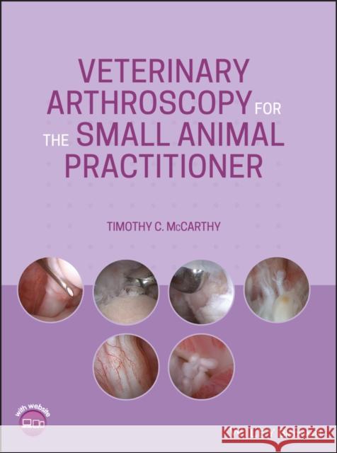
Veterinary Arthroscopy for the Small Animal Practitioner » książka


Veterinary Arthroscopy for the Small Animal Practitioner
ISBN-13: 9781119548973 / Angielski / Twarda / 2021 / 336 str.
Veterinary Arthroscopy for the Small Animal Practitioner
ISBN-13: 9781119548973 / Angielski / Twarda / 2021 / 336 str.
(netto: 765,71 VAT: 5%)
Najniższa cena z 30 dni: 794,18
ok. 30 dni roboczych.
Darmowa dostawa!
Preface xiAcknowledgments xiiiAbout the Companion Website xiv1 Introduction and Instrumentation 11.1 Introduction 11.2 Instrumentation and Equipment 31.2.1 Arthroscopes 31.2.2 Sheaths and Cannulas 51.2.2.1 Telescope Sheaths 51.2.2.2 Operative Cannulas 61.2.2.3 Egress Cannulas 81.2.3 Operative Hand Instruments 81.2.4 Power Instruments 121.2.4.1 Power Shavers 121.2.4.2 Radiofrequency/Electrocautery Instrumentation 141.2.5 Irrigation Fluid and Management Systems 151.2.5.1 Irrigation Fluids 151.2.5.2 Gravity Flow 161.2.5.3 Pressure Assisted Flow 161.2.5.4 Mechanical Arthroscopy Fluid Pumps 161.2.6 Video System Tower 171.2.6.1 Video Camera 181.2.6.2 Video Monitor 191.2.6.3 Light Source 191.2.6.4 Documentation Equipment 20References 202 General Technique 232.1 Anesthesia, Patient Support, and Pain Management 232.2 Postoperative Care 232.3 Patient Preparation, Positioning, and Operating Room Setup 242.3.1 Shoulder Joint 242.3.2 Elbow Joint 262.3.3 Radiocarpal Joint 282.3.4 Hip Joint 292.3.5 Stifle Joint 292.3.6 Tibiotarsal Joint 312.4 Portal Placement-General 31References 343 Shoulder Joint 363.1 Patient Preparation, Positioning, and Operating Room Setup 363.2 Portal Sites and Portal Placement 373.2.1 Telescope Portals 373.2.2 Operative Portals 393.2.3 Egress Portals 403.3 Nerves of Concern with Shoulder Joint Arthroscopy 403.4 Examination Protocol and Normal Arthroscopic Anatomy 413.5 Diseases of the Shoulder Diagnosed and Managed with Arthroscopy 473.5.1 Osteochondritis Dissecans (OCD) 473.5.1.1 OCD Lesion Removal and Management 593.5.2 Bicipital Tendon Injuries 733.5.3 Soft Tissue Injuries of the Shoulder with or Without Shoulder Instability 813.5.4 Ununited Caudal Glenoid Ossification Center (UCGOC) 953.5.5 Ununited Supraglenoid Tubercle (USGT) 1003.5.6 Arthroscopic-Assisted Intra-Articular Fracture Repair 1003.5.7 Arthroscopic Biopsy of Intra-Articular Neoplasia 1013.5.8 Glenoid Cartilage Defects 1023.5.9 Chondromalacia 1043.5.10 Infraspinatus Muscle Contracture 104References 1064 Arthroscopy of the Elbow Joint 1084.1 Patient Preparation, Positioning, and Operating Room Setup 1084.2 Portal Sites and Portal Placement 1094.2.1 Telescope Portals (Medial, Craniolateral, Caudomedial, and Caudal) 1094.2.2 Operative Portals (Craniomedial, Lateral, Craniolateral, and Caudal) 1114.2.3 Egress Portals 1134.3 Nerves of Concern with Elbow Joint Arthroscopy 1134.4 Examination Protocol and Normal Arthroscopic Anatomy 1144.5 Diseases of the Elbow Diagnosed and Managed with Arthroscopy 1224.5.1 Elbow Dysplasia 1224.5.2 Osteochondritis Dissecans (OCD) 1674.5.3 Ununited Anconeal Process (UAP) 1764.5.4 Degenerative Joint Disease (DJD) 1804.5.5 Assisted Intra-Articular Fracture Repair 1804.5.6 Biopsy of Intra-Articular Neoplasia 1814.5.7 Immune-Mediated Erosive Arthritis 1824.5.8 Incomplete Ossification of the Humeral Condyle (IOHC) 1824.5.9 Medial Enthesiopathy 183References 1845 Radiocarpal Joint 1875.1 Patient Preparation, Positioning, and Operating Room Setup 1875.2 Portal Sites and Portal Placement 1875.3 Nerves of Concern with Radiocarpal Joint Arthroscopy 1875.4 Examination Protocol and Normal Arthroscopic Anatomy 1885.5 Diseases of the Radiocarpal Joint Diagnosed and Managed with Arthroscopy 1895.5.1 Fractures 1895.5.2 Soft Tissue Injuries 1905.5.3 Immune-Mediated Erosive Arthritis 190References 1916 Hip Joint 1926.1 Patient Preparation, Positioning, and Operating Room Setup 1926.2 Portal Sites and Portal Placement 1926.3 Nerves of Concern with Hip Joint Arthroscopy 1936.4 Examination Protocol and Normal Arthroscopic Anatomy 1936.5 Diseases of the Hip Diagnosed and Managed with Arthroscopy 1966.5.1 Hip Dysplasia 1966.5.2 Arthroliths 2026.5.3 Soft Tissue Injuries of the Hip Joint 2036.5.4 Assisted Intra-Articular Fracture Repair 2056.5.5 Biopsy of Intra-Articular Neoplasia 2066.5.6 Aseptic Necrosis of the Femoral Head 206References 2067 Stifle Joint 2077.1 Patient Preparation, Positioning, and Operating Room Setup 2107.2 Portal Sites and Portal Placement 2107.2.1 Telescope Portal 2107.2.2 Operative Portals 2117.2.3 Egress Portal 2117.3 Nerves of Concern with Stifle Joint Arthroscopy 2137.4 Examination Protocol and Normal Arthroscopic Anatomy 2137.5 Diseases of the Stifle Joint Diagnosed and Managed with Arthroscopy 2217.5.1 Cranial Cruciate Ligament Injuries 2217.5.2 Caudal Cruciate Ligament Injuries 2587.5.3 Isolated Meniscal Injuries 2587.5.4 Osteochondritis Dissecans(OCD) 2597.5.5 Stifle Stabilization Failures 2647.5.6 TPLO Second Look 2647.5.7 Patellar Fracture Management 2667.5.8 Long Digital Extensor Tendon Injuries 2687.5.9 Popliteal Tendon Avulsion 2687.5.10 Intra-articular Neoplasia 2697.5.11 Patellar Luxation 2707.5.12 Degenerative Joint Disease, Chondromalacia, and Synovitis 2707.5.13 Discoid Meniscus 2737.5.14 Osteochondromatosis 274References 2748 Tibiotarsal Joint 2768.1 Patient Preparation, Positioning, and Operating Room Setup 2768.2 Portal Sites and Portal Placement 2768.2.1 Telescope Portals 2768.2.2 Operative Portals 2778.2.3 Egress Portal 2788.3 Nerves of Concern with Tibiotarsal Joint Arthroscopy 2788.4 Examination Protocol and Normal Arthroscopic Anatomy 2788.5 Diseases of the Tibiotarsal Joint Diagnosed and Managed with Arthroscopy 2798.5.1 Osteochondritis Dissecans (OCD) 2798.5.2 Intra-Articular Fracture Management 2868.5.3 Soft Tissue Injuries 2878.5.4 Immune-Mediated Erosive Arthritis 2878.5.5 Osteoarthritis 289References 2919 Problems and Complications 2929.1 Actual and Potential Complications of Arthroscopy 2929.1.1 Failure to Enter the Joint 2929.1.2 Articular Cartilage Damage 2929.1.3 Soft Tissue Damage 2969.1.4 Bone Fragment Displacement 2989.1.5 Operative Debris 2989.1.6 Red Out 2999.1.7 Peri-articular Fluid Accumulation 3009.1.8 Infection 3009.1.9 Vascular Injury 3019.1.10 Nerve Injury 3019.2 Instrument Damage 3019.2.1 Intra-articular Instrument Breakage 3019.2.2 Telescope Breakage 3029.3 Contraindications 3039.3.1 Patient Size 3039.3.2 Septic Arthritis 3039.3.3 Anesthesia Risk 303References 304Index 305
The authorTimothy C. McCarthy, DVM, PhD, Diplomate Emeritus, American College of Veterinary Surgeons, ACVS Founding Fellow, Minimally Invasive Surgery (Small Animal Orthopedics and Small Animal Soft Tissue) is an Educator at Veterinary Minimally Invasive Surgery Training (Vet MIST) in Beaverton, Oregon, USA.
1997-2026 DolnySlask.com Agencja Internetowa
KrainaKsiazek.PL - Księgarnia Internetowa


