• Wyszukiwanie zaawansowane
  • Kategorie
  • Kategorie BISAC
  • Książki na zamówienie
  • Promocje
  • Granty
  • Książka na prezent
  • Opinie
  • Pomoc
  • Załóż konto
  • Zaloguj się

Two-Dimensional Echocardiography in Infants and Children » książka

zaloguj się | załóż konto
Logo Krainaksiazek.pl

koszyk

konto

szukaj
topmenu
Księgarnia internetowa
Szukaj
Książki na zamówienie
Promocje
Granty
Książka na prezent
Moje konto
Pomoc
 
 
Wyszukiwanie zaawansowane
Pusty koszyk
Bezpłatna dostawa dla zamówień powyżej 20 złBezpłatna dostawa dla zamówień powyżej 20 zł

Kategorie główne

• Nauka
 [2948695]
• Literatura piękna
 [1824038]

  więcej...
• Turystyka
 [70868]
• Informatyka
 [151073]
• Komiksy
 [35227]
• Encyklopedie
 [23181]
• Dziecięca
 [621575]
• Hobby
 [138961]
• AudioBooki
 [1642]
• Literatura faktu
 [228651]
• Muzyka CD
 [371]
• Słowniki
 [2933]
• Inne
 [445341]
• Kalendarze
 [1243]
• Podręczniki
 [164416]
• Poradniki
 [479493]
• Religia
 [510449]
• Czasopisma
 [502]
• Sport
 [61384]
• Sztuka
 [243086]
• CD, DVD, Video
 [3417]
• Technologie
 [219673]
• Zdrowie
 [100865]
• Książkowe Klimaty
 [124]
• Zabawki
 [2168]
• Puzzle, gry
 [3372]
• Literatura w języku ukraińskim
 [260]
• Art. papiernicze i szkolne
 [7838]
Kategorie szczegółowe BISAC

Two-Dimensional Echocardiography in Infants and Children

ISBN-13: 9780898387780 / Angielski / Twarda / 1986 / 300 str.

J. P. Lintermans;Lintermans
Two-Dimensional Echocardiography in Infants and Children Lintermans, J. P. 9780898387780 Springer - książkaWidoczna okładka, to zdjęcie poglądowe, a rzeczywista szata graficzna może różnić się od prezentowanej.

Two-Dimensional Echocardiography in Infants and Children

ISBN-13: 9780898387780 / Angielski / Twarda / 1986 / 300 str.

J. P. Lintermans;Lintermans
cena 803,21
(netto: 764,96 VAT:  5%)

Najniższa cena z 30 dni: 771,08
Termin realizacji zamówienia:
ok. 22 dni roboczych.

Darmowa dostawa!

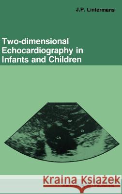
In 1981 Dr Jean Lintermans published, with Dr van Dorp, a superb vol- ume, 'Differential Diagnosis in Pediatric Echocardiography'. It was 'state of the art' with a unique organization, starting with M-mode echocardiograph- ic findings rather than the disease category, and included a segmental approach to diagnosis, and concluded with an invaluable section on normal echocardiographic values. This volume was a great help to many of us in pediatric cardiology who were discovering the great clinical value of the noninvasive approach to diagnosis. This gave us a 'running start' for our own entry into the field. At that time, the 2 D or sector echocardiogram was finding increasing usefulness and there were several illustrations of this tech- nique, integrated into that volume. Since 1980, the field of 2 D echocardiography has grown enormously in its usefulness, to the point that it has reduced the need for invasive studies, and has enhanced the precision of invasive studies when required. It is now time for a systematic and thorough approach to this field, and I am delighted that Jean Lintermans has provided us with this book. The pictures are uniformly superb and are very well labelled. The organization is cen- tered around diagnostic categories, but the same attention to detail is pre- sent that made the first volume so useful. I particularly value the extensive documentation of diagnostic findings, with numerous literature citations.

Kategorie:
Nauka, Medycyna
Kategorie BISAC:
Medical > Pediatrics
Medical > Kardiologia
Wydawca:
Springer
Język:
Angielski
ISBN-13:
9780898387780
Rok wydania:
1986
Wydanie:
1986
Ilość stron:
300
Waga:
0.64 kg
Wymiary:
24.18 x 16.87 x 2.36
Oprawa:
Twarda
Wolumenów:
01
Dodatkowe informacje:
Bibliografia
Wydanie ilustrowane

1. Left-to-right shunts.- 1.1. Congenital left-to-right shunts.- 1.1.1. Atrial septal defects.- 1.1.2. Ventricular septal defects.- 1.1.3. Atrioventricular canal defects.- 1.1.4. Common atrium.- 1.1.5. Patent ductus arteriosus.- 1.1.6. Aorto-pulmonary window.- 1.1.7. Intracranial arteriovenous malformation.- 1.2. Acquired left-to-right shunts.- 1.2.1. Atrial septal defect.- 1.2.2. Aortopulmonary shunts.- 2. Conotruncal abnormalities.- 2.1. Tetralogy of Fallot.- 2.2. Persistent truncus arteriosus.- 2.3. Pulmonary atresia with ventricular septal defect.- 2.4. Double outlet right ventricle.- 3. Left ventricular outflow obstruction.- 3.1. Aortic valve stenosis.- 3.2. Subvalvular aortic stenosis.- 3.3. Supravalvar aortic stenosis.- 3.4. Coarctation of the aorta.- 3.5. Interruption of the aortic arch.- 3.6. Double aortic arch.- 4. Right ventricular outflow obstruction.- 4.1. Congenital right ventricular outflow obstruction.- 4.1.1. Pulmonary valve stenosis.- 4.1.2. Right ventricular outflow tract obstruction.- 4.1.3. Peripheral pulmonary artery obstruction.- 4.2. Acquired right ventricular outflow obstruction.- 4.2.1. Pulmonary artery banding.- 4.2.2. Right ventricular outflow tract obstruction.- 5. Left ventricular inflow obstruction.- 5.1. Congenital left ventricular inflow obstruction.- 5.1.1. Mitral valve stenosis.- 5.1.2. Cor triatriatum.- 5.1.3. Supravalvar mitral ring.- 5.1.4. Pulmonary vein stenosis.- 5.2. Acquired left ventricular inflow obstruction.- 5.2.1. Rheumatic mitral valve stenosis.- 5.2.2. After Mustard/Senning operation.- 6. Right ventricular inflow obstruction.- 6.1. Congenital right ventricular inflow obstruction.- 6.1.1. Tricuspid atresia.- 6.1.2. Tricuspid stenosis.- 6.2. Acquired right ventricular inflow obstruction.- 6.2.1. Tricuspid stenosis.- 7. Assessment of valvular regurgitation and valvular prolapse.- 7.1. Mitral valve.- 7.1.1. Mitral valve prolapse.- 7.1.2. Ruptured chordae tendineae.- 7.1.3. Mitral valve regurgitation.- 7.2. Tricuspid valve.- 7.2.1. Tricuspid valve prolapse.- 7.2.2. Tricuspid valve regurgitation.- 7.3. Aortic valve.- 7.3.1. Aortic valve prolapse.- 7.3.2. Aortic valve regurgitation.- 8. Transposition of the great arteries.- 8.1. d-Transposition of the great arteries.- 8.2. 1-Transposition of the great arteries, with ventricular inversion.- 8.3. d-Transposition of the great arteries after hemodynamic correction.- 8.4. d-Transposition of the great arteries after anatomic correction.- 9. Total anomalous pulmonary venous return.- 9.1. Supracardiac TAPVR.- 9.2. Cardiac TAPVR.- 9.3. Infradiaphragmatic TAPVR.- 10. Ebstein’s anomaly of the tricuspid valve.- 11. Hypoplastic heart syndromes.- 11.1. Hypoplastic left heart syndrome.- 11.2. Pulmonary valve atresia, including the hypoplastic right heart syndrome.- 11.3. Overriding and straddling atrioventricular (AV) valves.- 11.4. Single ventricle.- 11.5. Uhl’s anomaly.- 12. Myocardial diseases.- 12.1. Hypertrophic cardiomyopathy (HCM).- 12.2. Congestive cardiomyopathies.- 12.3. Double chambered right ventricle.- 13. Pericardial and pleural affections.- 13.1. Pericardial effusion.- 13.2. Cardiac tamponade.- 13.3. Constructive pericarditis.- 13.4. Pleural effusion.- 14. Tumors and thrombi.- 14.1. Cardiac tumors and thrombi.- 14.2. Mediastinal tumors.- 15. Aneurysms.- 15.1. Ventricular wall aneurysm.- 15.2. Aneurysm of the ventricular septum.- 15.3. Atrial septal aneurysm.- 15.4. Sinus of Valsalva aneurysm and related pathology.- 15.4.1. Sinus of Valsalva aneurysm.- 15.4.2. Aorto-left ventricular tunnel.- 15.4.3. Ventricular septal defect with aortic regurgitation.- 16. Endocarditis.- 16.1. Bacterial endocarditis.- 16.2. Vegetative lesions.- 16.3. Complications or hemodynamic sequels.- 16.3.1. Flail valve leaflets.- 16.3.2. Annular erosion with abcess formation.- 16.3.3. Left ventricular pseudoaneurysms.- 16.3.4. Mitral leaflet aneurysms.- 16.3.5. Leaflet perforation.- 17. Foreign bodies.- 17.1. Patches.- 17.2. Conduits.- 17.3. Ventriculo-cardiac shunts.- 17.4. Pacemaker wires.- 18. Not commonly visualized cardiovascular structures.- 18.1. Left superior vena cava (LSVC) and coronary sinus.- 18.2. Persistence of right sinus venosus valve.- 18.3. False tendons.- 19. Malformation syndromes with their typical cardiovascular abnormalities and corresponding ultrasonic features.- 19. 1. Trisomy 21 (DOWN) syndrome.- 19. 2. Gonadal agenesis or Turner syndrome.- 19. 3. Noonan syndrome.- 19. 4. Infants of diabetic mothers.- 19. 5. Rubella syndrome.- 19. 6. Tuberous sclerosis.- 19. 7. Williams-Beuren syndrome (supravalvar aortic stenosis with elf-like facies.- 19. 8. Marfan syndrome.- 19. 9. Holt-Oram syndrome.- 19.10. Pompe’s disease (type 2 glycogen storage disease).- 19.11. Multiple lentigines or leopard syndrome.- 19.12. Intrahepatic biliary atresia with peripheral pulmonary artery stenosis or Alagille syndrome.- 19.13. DiGeorge syndrome.- 19.14. Ellis-Van Creveld syndrome.- 19.15. Mucocutaneous lymph node syndrome or Kawasaki disease.- 20. Segmental approach to the diagnosis of congenital malformation and malposition.- 20.1. The atrial situs.- 20.2. The pattern of systematic venous drainage.- 20.3. The pattern of pulmonary venous drainage.- 20.4. Position of cardiac apex.- 20.5. Definition of ventricular morphology and location.- 20.6. Atrioventricular (AV) connections.- 20.7. Identification of great arteries.- 20.8. Assessment of ventriculo-arterial connections.- 20.9. Detection of the aortic arch.- Index of Subjects.


Udostępnij

Facebook - konto krainaksiazek.pl


Opinie o Krainaksiazek.pl na Opineo.pl

Partner Mybenefit

Krainaksiazek.pl w programie rzetelna firma Krainaksiaze.pl - płatności przez paypal

Czytaj nas na:

Facebook - krainaksiazek.pl
  • książki na zamówienie
  • granty
  • książka na prezent
  • kontakt
  • pomoc
  • opinie
  • regulamin
  • polityka prywatności

Zobacz:

  • Księgarnia czeska

  • Wydawnictwo Książkowe Klimaty

1997-2026 DolnySlask.com Agencja Internetowa

© 1997-2022 krainaksiazek.pl
     
KONTAKT | REGULAMIN | POLITYKA PRYWATNOŚCI | USTAWIENIA PRYWATNOŚCI
Zobacz: Księgarnia Czeska | Wydawnictwo Książkowe Klimaty | Mapa strony | Lista autorów
KrainaKsiazek.PL - Księgarnia Internetowa
Polityka prywatnosci - link
Krainaksiazek.pl - płatnośc Przelewy24
Przechowalnia Przechowalnia